Session Information
Session Type: Abstract Submissions (ACR)
Background/Purpose: Hypoxia, leukocyte infiltration and dysfunctional vascularity play a key role in the pathogenesis of inflammatory arthritis (IA). We examine the relationship of metabolic turnover with synovial vascularity / invasion in IA patients.
Methods: RA and PsA patients with active knee joint synovitis were recruited and assessed clinically prior to contiguous MR and PET/CT imaging followed by arthroscopy, biopsy and pO2 measurement pre- and post-TNFi therapy. Imaging protocols were standardised for MRI, and Fluorodeoxyglucose (18FDG) PET and image-data intensity resolution was optimised for synovial tissue. Cell-specific markers (CD3, CD4, CD8, CD68), blood vessel maturity and metabolic activity (GAPDH) were quantified by immunohistologic analysis and dual-immunofluorescence. Reduction of DAS28 ≥1.2 indicated a good clinical response. Mann Whitney U and Wilcoxon test were used to compare continuous variables and Spearman’s Rho was used to assess correlations. p<0.05 was considered statistically significant. SPSS version 18 was employed.
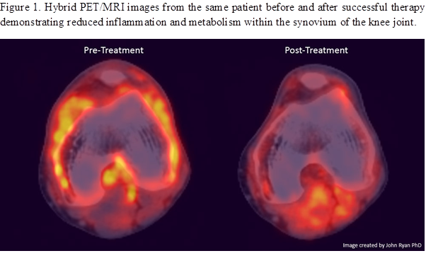
Results: 21 patients, RA(n=9) and PsA(n=12), mean (age 45±16, DAS 28, 4.6±1.2 and, gender 6F:15M) were recruited. At baseline in vivo synovial pO2 levels inversely correlated with PET (r=-0.6, p=0.03), DAS28 (r=-0.5, p=0.05) and was inversely associated with macroscopic synovitis. Furthermore, MRI correlated with macroscopic synovitis (r=0.5, p=0.04) and sub-linining CD68 expression (r=0.7, p=0.001). Lining layer and sub-lining GAPDH expression inversely correlated with synovial pO2 (r=0.8, p=0.03) pre/post TNFi. Biologic responders showed significant reduction in PET quantification (p=0.018) which was paralleled by corresponding decreases in MRI (p=0.012), macroscopic synovitis (p=0.005), vascularity (p=0.005) and CD68sl expression (p=0.01) and an increase in pO2 levels from 22mmHG to 37 mmHG. In contrast PET quantification in non-responders was not associated with significant reductions in MRI or macroscopic/microscopic measures of disease. Furthermore pO2 levels in non-responders were similar pre/post therapy (22mmHG vs 25mmHG). Figure 1 shows representative PET/MRI hybrid images in a responder patient pre/post TNFi demonstrating close association between metabolic turnover and site of inflammation.
Conclusion: This is the first study to show synovial metabolic activity and inflammation quantified by MR/PET are associated with in vivo hypoxia, cellular and molecular biomarkers in RA and PsA patients in response to biologic therapy. MR/PET may represent an important new imaging modality in assessing response to biologic therapy.
Disclosure:
L. C. Harty,
None;
J. Ryan,
None;
C. T. Ng,
None;
M. Biniecka,
None;
A. Kennedy,
None;
E. J. Heffernan,
None;
U. Fearon,
None;
D. J. Veale,
Roche Pharmaceuticals,
5,
Pfizer Inc, MSD, Bayer,
5,
Pfizer Inc, MSD, Medimmune,
8.
« Back to 2012 ACR/ARHP Annual Meeting
ACR Meeting Abstracts - https://acrabstracts.org/abstract/in-vivo-synovial-oxygen-levels-are-inversely-related-to-metabolic-turnover-and-disease-activity-in-rheumatoid-and-psoriatic-arthritis-biologic-responders/

