ACR Meeting Abstracts

ACR Meeting Abstracts

  • Meetings
    • ACR Convergence 2024
    • ACR Convergence 2023
    • 2023 ACR/ARP PRSYM
    • ACR Convergence 2022
    • ACR Convergence 2021
    • ACR Convergence 2020
    • 2020 ACR/ARP PRSYM
    • 2019 ACR/ARP Annual Meeting
    • 2018-2009 Meetings
    • Download Abstracts
  • Keyword Index
  • Advanced Search
  • Your Favorites
    • Favorites
    • Login
    • View and print all favorites
    • Clear all your favorites
  • ACR Meetings

Abstract Number: 1557

Visualization of Dorsal Root Ganglionitis with Three-Tesla Magnetic Resonance Neurography in Sensory Ataxic Neuropathy Associated with Sjögren’s Syndrome

Takeshi Yoshida1, Takeshi Sueyoshi2, Mitsuyo Kinjo3, Shugo Suwazono4 and Hiroyuki Nodera5, 1Rheumatology, Chikamori Hospital, Kochi, Japan, 2Radiology, Minei Daiichi Hospital, Urasoe, Japan, 3Rheumatology, Okinawa Chubu Hospital, Uruma, Japan, 4Brain-Nerve-Muscle Research Center, National Hospital Organization Okinawa Hospital, Uruma, Japan, 5Neurology, Tokushima University School of Medicine, Tokushima, Japan

Meeting: 2018 ACR/ARHP Annual Meeting

Keywords: MRI, neuroimaging and neuropathy, Sjogren's syndrome

  • Tweet
  • Click to email a link to a friend (Opens in new window) Email
  • Click to print (Opens in new window) Print
Session Information

Date: Monday, October 22, 2018

Title: Sjögren's Syndrome – Basic and Clinical Science Poster

Session Type: ACR Poster Session B

Session Time: 9:00AM-11:00AM

Background/Purpose: Sjögrenfs syndrome (SS)-associated neuropathy manifests as various forms of neuropathy, including sensory ataxic neuropathy (SAN). Dorsal root ganglionitis, pathologically defined as the lymphocytic infiltration of the dorsal root ganglion (DRG), causes SAN. This study aimed to determine whether 3-Tesla magnetic resonance neurography (3T-MRN) is useful for detecting abnormalities of DRGs, and making diagnoses in patients with SS-SAN.

Methods: We conducted a retrospective chart review from 2015 to 2017 and enrolled 3 patients with SS-SAN fulfilling American–European Consensus Group classification criteria. We evaluated with a 3T MRI scanner and used the coronal short-tau inversion recovery (STIR) technique because of its advantages in depicting a symmetric normal hyperintensity of DRGs and nerves. On measuring a pair of DRGs, regions of interest (ROI) were manually drawn by using ImageJ (http://rsb.info.nih.gov/ij/). The area, median SI, and transverse diameter of each DRGs were measured and compared with those of the corresponding proximal nerve roots. The SI ratio (SI within a DRG/SI within a proximal nerve root) and diameter ratio (transverse diameter of a DRG/width at the middle point of a proximal nerve root) were subsequently calculated. For each nerve root level, data from SS patients were compared with those of age- and sex-matched non-neuropathy controls (20 DRGs from 10 cases). All statistical analyses were performed with EZR, a graphical user interface for R (The R Foundation for Statistical Computing, Vienna, Austria). The Mann–Whitney U test was applied for the statistical analysis.

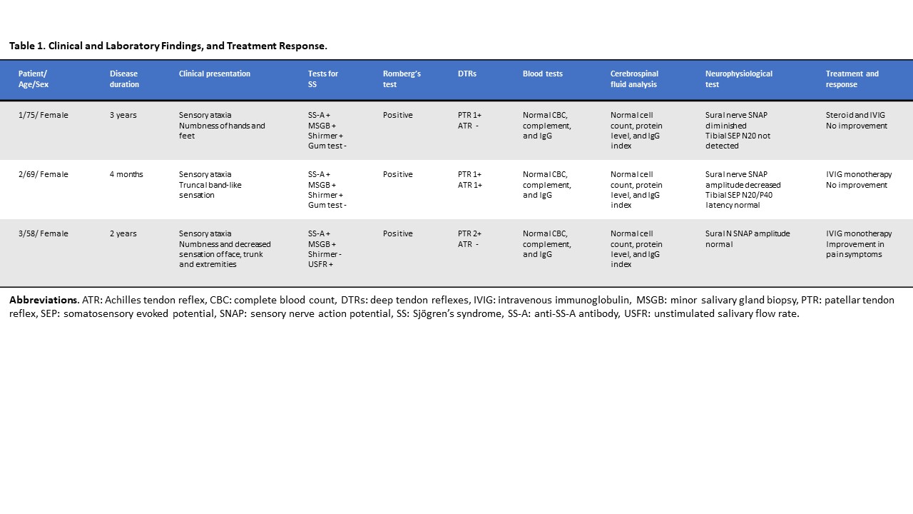
Results: Table 1 summarizes the clinical data of the 3 patients. These SS-SAN patients presented with sensory ataxia with positive Rombergfs test and also had various painful symptoms. All 3 patients showed normal motor strength. Compared with the control data, all DRGs in SS patients were numerically smaller and had a lower SI through the L2-S1 level. Moreover, the area, diameter ratio, and SI ratio were significantly reduced in L3-L4 DRGs (Table 2).

Conclusion: We revealed that the DRGs in patients with SS-SAN were atrophic and showed a decreased SI than those in the controls. We suggest the visualization of DRGs by 3T-MRN as a potentially useful imaging biomarker for the clinical diagnosis, as well as the assessment of pathophysiological mechanisms. DRG atrophy and decreased signal intensity may reflect the sensory neuronal loss caused by chronic lymphocytic inflammation in DRGs.


Disclosure: T. Yoshida, None; T. Sueyoshi, None; M. Kinjo, None; S. Suwazono, None; H. Nodera, None.

To cite this abstract in AMA style:

Yoshida T, Sueyoshi T, Kinjo M, Suwazono S, Nodera H. Visualization of Dorsal Root Ganglionitis with Three-Tesla Magnetic Resonance Neurography in Sensory Ataxic Neuropathy Associated with Sjögren’s Syndrome [abstract]. Arthritis Rheumatol. 2018; 70 (suppl 9). https://acrabstracts.org/abstract/visualization-of-dorsal-root-ganglionitis-with-three-tesla-magnetic-resonance-neurography-in-sensory-ataxic-neuropathy-associated-with-sjogrens-syndrome/. Accessed .
  • Tweet
  • Click to email a link to a friend (Opens in new window) Email
  • Click to print (Opens in new window) Print

« Back to 2018 ACR/ARHP Annual Meeting

ACR Meeting Abstracts - https://acrabstracts.org/abstract/visualization-of-dorsal-root-ganglionitis-with-three-tesla-magnetic-resonance-neurography-in-sensory-ataxic-neuropathy-associated-with-sjogrens-syndrome/

Advanced Search

Your Favorites

You can save and print a list of your favorite abstracts during your browser session by clicking the “Favorite” button at the bottom of any abstract. View your favorites »

All abstracts accepted to ACR Convergence are under media embargo once the ACR has notified presenters of their abstract’s acceptance. They may be presented at other meetings or published as manuscripts after this time but should not be discussed in non-scholarly venues or outlets. The following embargo policies are strictly enforced by the ACR.

Accepted abstracts are made available to the public online in advance of the meeting and are published in a special online supplement of our scientific journal, Arthritis & Rheumatology. Information contained in those abstracts may not be released until the abstracts appear online. In an exception to the media embargo, academic institutions, private organizations, and companies with products whose value may be influenced by information contained in an abstract may issue a press release to coincide with the availability of an ACR abstract on the ACR website. However, the ACR continues to require that information that goes beyond that contained in the abstract (e.g., discussion of the abstract done as part of editorial news coverage) is under media embargo until 10:00 AM ET on November 14, 2024. Journalists with access to embargoed information cannot release articles or editorial news coverage before this time. Editorial news coverage is considered original articles/videos developed by employed journalists to report facts, commentary, and subject matter expert quotes in a narrative form using a variety of sources (e.g., research, announcements, press releases, events, etc.).

Violation of this policy may result in the abstract being withdrawn from the meeting and other measures deemed appropriate. Authors are responsible for notifying colleagues, institutions, communications firms, and all other stakeholders related to the development or promotion of the abstract about this policy. If you have questions about the ACR abstract embargo policy, please contact ACR abstracts staff at [email protected].

Wiley

  • Online Journal
  • Privacy Policy
  • Permissions Policies
  • Cookie Preferences

© Copyright 2025 American College of Rheumatology