ACR Meeting Abstracts

ACR Meeting Abstracts

  • Meetings
    • ACR Convergence 2024
    • ACR Convergence 2023
    • 2023 ACR/ARP PRSYM
    • ACR Convergence 2022
    • ACR Convergence 2021
    • ACR Convergence 2020
    • 2020 ACR/ARP PRSYM
    • 2019 ACR/ARP Annual Meeting
    • 2018-2009 Meetings
    • Download Abstracts
  • Keyword Index
  • Advanced Search
  • Your Favorites
    • Favorites
    • Login
    • View and print all favorites
    • Clear all your favorites
  • ACR Meetings

Abstract Number: 2683

Visual and Quantitative Assessment of Cranial Arteries on FDG-PET/CT Can Reliably Diagnose Cranial Giant Cell Arteritis

Pieter Nienhuis1, Riemer Slart 2 and Elisabeth Brouwer 3, 1Dept. of Rheumatology and Clinical Immunology - University Medical Center Groningen, Groningen, Groningen, Netherlands, 2Dept. of Nuclear Medicine and Molecular Imaging - University Medical Center Groningen, Groningen, Groningen, Netherlands, 3Department of Rheumatology and Clinical Immunology, University of Groningen, University Medical Center Groningen, Groningen, Netherlands

Meeting: 2019 ACR/ARP Annual Meeting

Keywords: 18FDG PET/CT scan and diagnostic imaging, giant cell arteritis

  • Tweet
  • Click to email a link to a friend (Opens in new window) Email
  • Click to print (Opens in new window) Print
Session Information

Date: Tuesday, November 12, 2019

Title: Vasculitis – Non-ANCA-Associated & Related Disorders Poster III: Giant Cell Arteritis

Session Type: Poster Session (Tuesday)

Session Time: 9:00AM-11:00AM

Background/Purpose: Assessing cranial artery inflammation plays an important role in the diagnosis of cranial giant cell arteritis (GCA). Although an established tool for assessing large vessel vasculitis, fluorine-18-deoxyglucose (FDG) positron emission tomography (PET)/CT imaging is not recommended for detecting cranial artery inflammation. Assessing FDG uptake in the cranial arteries may increase the diagnostic value of FDG-PET/CT for GCA and may provide more detailed information regarding involved arteries and extent of the disease. It may thus provide a viable alternative for Duplex ultrasonography (US) and temporal artery biopsy (TAB) as a first line investigation in GCA.
This study aimed to evaluate the diagnostic accuracy of FDG-PET/CT for the detection of cranial artery inflammation in GCA.

Methods: This retrospective case control study included glucocorticoid-naive TAB positive GCA patients who underwent FDG-PET/CT as part of the diagnostic work-up. Controls were age- and sex-matched melanoma patients in remission with FDG-PET/CT after 6 months follow-up.

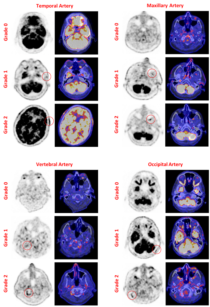
To attain high quality PET images on the Siemens Biograph 64 PET/CT, optimal PET image acquisition and reconstruction parameters were determined and applied to all images. Visual assessment by an experienced nuclear medicine physician was based on uptake intensity compared to surrounding tissue on a 0-2 scale (Figure 1). Additionally, FDG uptake in the cranial arteries was quantified by measuring the maximum standardized uptake value (SUVmax) in the target arteries. The temporal arteries (TA), maxillary arteries (MA), vertebral arteries (VA), and occipital arteries (OA) were assessed bilaterally.

Student t and chi-squared tests were used for evaluation of the visual assessment. Receiver operating characteristics (ROC) analysis was performed for evaluation of the quantitative assessment. Cohen’s kappa was used to assess agreement of the visual and quantitative assessments.

Results: A total of 24 GCA patients and 24 controls were included. The mean age for both groups was 71 years (SD: 8) and 12/24 were women. Large vessel involvement was seen on PET in 15/24 patients. Duplex US of the temporal artery was performed in 15/24 patients, of which 9 were positive. Median ESR and CRP were 73 (CI95%: 53; 104) mm/h and 73 (CI95%: 37; 110) mg/L, respectively.

Visual assessment revealed a sensitivity of 83% (CI95%: 64; 93) and a specificity of 75% (CI95%: 55; 88) for GCA denoted by a score of ≥1 (uptake higher than background). Receiver-operating characteristic (ROC) analysis of the quantitative assessment revealed an AUC of 0.88 (CI95%­­=0.77-0.99) resulting in a sensitivity of 79% (CI95%: 60; 91) and a specificity of 92% (CI95%: 74; 99) for a SUVmax threshold value of 5.00 (Table 1). The agreement between the two assessment methods was 77% (kappa 0.55).

Conclusion: Using optimal scan data reconstruction, visual and quantitative FDG-PET/CT assessment methods can reliably diagnose GCA based on cranial artery inflammation. Extending the use of FDG-PET/CT to the cranial arteries may improve the diagnostic accuracy for GCA. Moderate agreement between visual and quantitative assessment methods suggests that diagnostic accuracy may be improved when combining both methods.

Figure 1. Visual assessment of cranial artery inflammation on FDG-PET/CT. Temporal Artery -TA-, Maxillary Artery -MA-, Vertebral Artery -VA-, and Occipital Artery -OA- were visually scored from 0-2. Scoring definition: 0: no uptake above surrounding tissue; 1: uptake just above surrounding tissue; 2: uptake significantly above surrounding tissue. The red circle denotes the visually determined area of increased uptake.

Table 1. Cross tabulation of the results of the visual and the quantitative assessments of FDG uptake in the cranial arteries. Visual scoring definition: 0: no uptake above surrounding tissue; 1: uptake just above surrounding tissue; 2: uptake significantly above surrounding tissue. TA, temporal artery; MA, maxillary artery; VA, vertebral artery; OA, occipital artery; SUVmax, maximum standardized uptake value; CI95%, 95% confidence interval.


Disclosure: P. Nienhuis, None; R. Slart, None; E. Brouwer, Roche, 5, 8.

To cite this abstract in AMA style:

Nienhuis P, Slart R, Brouwer E. Visual and Quantitative Assessment of Cranial Arteries on FDG-PET/CT Can Reliably Diagnose Cranial Giant Cell Arteritis [abstract]. Arthritis Rheumatol. 2019; 71 (suppl 10). https://acrabstracts.org/abstract/visual-and-quantitative-assessment-of-cranial-arteries-on-fdg-pet-ct-can-reliably-diagnose-cranial-giant-cell-arteritis/. Accessed .
  • Tweet
  • Click to email a link to a friend (Opens in new window) Email
  • Click to print (Opens in new window) Print

« Back to 2019 ACR/ARP Annual Meeting

ACR Meeting Abstracts - https://acrabstracts.org/abstract/visual-and-quantitative-assessment-of-cranial-arteries-on-fdg-pet-ct-can-reliably-diagnose-cranial-giant-cell-arteritis/

Advanced Search

Your Favorites

You can save and print a list of your favorite abstracts during your browser session by clicking the “Favorite” button at the bottom of any abstract. View your favorites »

All abstracts accepted to ACR Convergence are under media embargo once the ACR has notified presenters of their abstract’s acceptance. They may be presented at other meetings or published as manuscripts after this time but should not be discussed in non-scholarly venues or outlets. The following embargo policies are strictly enforced by the ACR.

Accepted abstracts are made available to the public online in advance of the meeting and are published in a special online supplement of our scientific journal, Arthritis & Rheumatology. Information contained in those abstracts may not be released until the abstracts appear online. In an exception to the media embargo, academic institutions, private organizations, and companies with products whose value may be influenced by information contained in an abstract may issue a press release to coincide with the availability of an ACR abstract on the ACR website. However, the ACR continues to require that information that goes beyond that contained in the abstract (e.g., discussion of the abstract done as part of editorial news coverage) is under media embargo until 10:00 AM ET on November 14, 2024. Journalists with access to embargoed information cannot release articles or editorial news coverage before this time. Editorial news coverage is considered original articles/videos developed by employed journalists to report facts, commentary, and subject matter expert quotes in a narrative form using a variety of sources (e.g., research, announcements, press releases, events, etc.).

Violation of this policy may result in the abstract being withdrawn from the meeting and other measures deemed appropriate. Authors are responsible for notifying colleagues, institutions, communications firms, and all other stakeholders related to the development or promotion of the abstract about this policy. If you have questions about the ACR abstract embargo policy, please contact ACR abstracts staff at [email protected].

Wiley

  • Online Journal
  • Privacy Policy
  • Permissions Policies
  • Cookie Preferences

© Copyright 2025 American College of Rheumatology