ACR Meeting Abstracts

ACR Meeting Abstracts

  • Meetings
    • ACR Convergence 2024
    • ACR Convergence 2023
    • 2023 ACR/ARP PRSYM
    • ACR Convergence 2022
    • ACR Convergence 2021
    • ACR Convergence 2020
    • 2020 ACR/ARP PRSYM
    • 2019 ACR/ARP Annual Meeting
    • 2018-2009 Meetings
    • Download Abstracts
  • Keyword Index
  • Advanced Search
  • Your Favorites
    • Favorites
    • Login
    • View and print all favorites
    • Clear all your favorites
  • ACR Meetings

Abstract Number: 2448

UV Light Exposure Induces a Type I Interferon Dependent Activation and Migration of Inflammatory Dendritic Cells to Local Lymph Nodes

Xizhang Sun, Jaime Chao, Michael Gerner and Keith Elkon, University of Washington, Seattle, WA

Meeting: ACR Convergence 2023

Keywords: Cutaneous, Dendritic cells, interferon

  • Tweet
  • Click to email a link to a friend (Opens in new window) Email
  • Click to print (Opens in new window) Print
Session Information

Date: Tuesday, November 14, 2023

Title: Abstracts: SLE – Animal Models

Session Type: Abstract Session

Session Time: 2:00PM-3:30PM

Background/Purpose: Photosensitivity occurs in ~ 75% of lupus patients. Although ultraviolet (UV) light stimulates Type I interferon (IFN-I) in the skin, why lupus patients are sensitive to UV skin injury and how systemic immune responses are generated are poorly understood. Following skin inflammation in other contexts, DC migrate to draining lymph nodes (dLN) and activate adaptive immune cells. In virus infections that induce Type I interferon (IFN-I), a unique inflammatory DC subset (infl-cDC2) that are potent antigen presenting cells are generated. To explore the links between UV mediated skin inflammation and innate responses, we examined myeloid cell migration, including DC subsets (cDC1, cDC2, infl-cDC2) to dLNs in normal and a lupus prone mouse strain. We also examined the influence of Type-I IFN in migratory DC differentiation.

Methods: Mice were females aged 8-12 weeks of the strains C57BL/6 (B6); KiKR (photoactivatable); interferon receptor deficient (Ifnar KO); and Trex1 mutant mice (knock in of the lupus mutant allele, D18N, Trex1m). Mice were exposed to a single dose of UV (500mJ/cm2) on the dorsal skin. In some experiments, the skin was painted with TRITC prior to UV to determine the origin of cells in the dLN. Brachial and axillary dLN were harvested at 1- or 2-days post UV exposure. Flow cytometry using the Symphony A3 cytometer identified myeloid cells as described previously. Statistical significance between groups was determined by Student’s t-test.

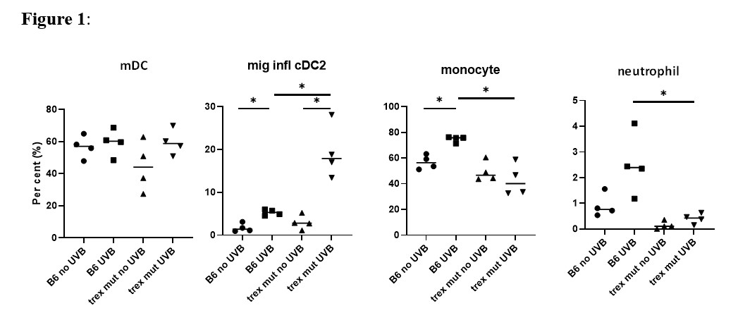
Results: Skin painted with TRITC confirmed that skin derived DC migrated to dLN after UV. When compared to non-UV exposed mice and control non-draining LN, UV caused an increase in TRITC+CD64+ and TRITC+ Ly6Ghi (neutrophils) cells in dLN. UV of KiKR mouse skin revealed that both cDC1 and cDC2 cells migrated to dLN following UV. Higher numbers of neutrophils as well as CD64+ myeloid cells, that included monocytes as well as infl-cDC2 (CD26+MAR-1+), were detected in the dLN after UV, compared to non-UV exposed control mice (Fig.1). We observed a significant reduction of infl-cDC2 cells in Ifnar KO mice indicating that their development was IFN-I dependent. Finally, we quantified myeloid cell populations in the dLN of lupus-prone Trex1m mice. Although UV did not increase cDC1 nor cDC2 in dLN, we observed a significant increase in the infl-cDC2 in the dLN in this lupus prone strain (Fig.1). Curiously, monocyte and neutrophil migration to dLN were reduced in Trex1m mice compared to B6 after UV.

Conclusion: We conclude that exposure of normal skin to UV light causes migration of many different types of myeloid cells to dLN, including skin-derived migratory DC, neutrophils, and monocytes. A subset of CD64+ infl-cDC2 is also induced and is dependent on UV-induced IFN-I. Of interest, Trex1m mice exhibit altered myeloid cell responses in the dLNs, including increased representation of infl-DCs and reduced neutrophil and monocyte cellularity. Overall, these findings show marked and non-equivalent IFN-I dependent effects of UV on myeloid cell responses in the dLN in wild type vs lupus-prone mice, implicating a link between IFN-I and the development of APCs that promote the adaptive immune response.

Supporting image 1

Fig 1. Percentage of cells in the draining Lymph Nodes (dLN). B6 or Trex1 mutant (mut) mice were exposed to 500 mJ/cm2 UV light on the dorsum. 48 hours after UV exposure, the brachial and axillary LN were harvested. The dissociated cells were analyzed by flow cytometry using 15 markers to distinguish different myeloid population on the Symphony A3 cytometer. The percentage of each cell type was calculated and comparisons made by Student’s t test. mDC = migratory DC. * p<0.05.


Disclosures: X. Sun: None; J. Chao: None; M. Gerner: None; K. Elkon: None.

To cite this abstract in AMA style:

Sun X, Chao J, Gerner M, Elkon K. UV Light Exposure Induces a Type I Interferon Dependent Activation and Migration of Inflammatory Dendritic Cells to Local Lymph Nodes [abstract]. Arthritis Rheumatol. 2023; 75 (suppl 9). https://acrabstracts.org/abstract/uv-light-exposure-induces-a-type-i-interferon-dependent-activation-and-migration-of-inflammatory-dendritic-cells-to-local-lymph-nodes/. Accessed .
  • Tweet
  • Click to email a link to a friend (Opens in new window) Email
  • Click to print (Opens in new window) Print

« Back to ACR Convergence 2023

ACR Meeting Abstracts - https://acrabstracts.org/abstract/uv-light-exposure-induces-a-type-i-interferon-dependent-activation-and-migration-of-inflammatory-dendritic-cells-to-local-lymph-nodes/

Advanced Search

Your Favorites

You can save and print a list of your favorite abstracts during your browser session by clicking the “Favorite” button at the bottom of any abstract. View your favorites »

All abstracts accepted to ACR Convergence are under media embargo once the ACR has notified presenters of their abstract’s acceptance. They may be presented at other meetings or published as manuscripts after this time but should not be discussed in non-scholarly venues or outlets. The following embargo policies are strictly enforced by the ACR.

Accepted abstracts are made available to the public online in advance of the meeting and are published in a special online supplement of our scientific journal, Arthritis & Rheumatology. Information contained in those abstracts may not be released until the abstracts appear online. In an exception to the media embargo, academic institutions, private organizations, and companies with products whose value may be influenced by information contained in an abstract may issue a press release to coincide with the availability of an ACR abstract on the ACR website. However, the ACR continues to require that information that goes beyond that contained in the abstract (e.g., discussion of the abstract done as part of editorial news coverage) is under media embargo until 10:00 AM ET on November 14, 2024. Journalists with access to embargoed information cannot release articles or editorial news coverage before this time. Editorial news coverage is considered original articles/videos developed by employed journalists to report facts, commentary, and subject matter expert quotes in a narrative form using a variety of sources (e.g., research, announcements, press releases, events, etc.).

Violation of this policy may result in the abstract being withdrawn from the meeting and other measures deemed appropriate. Authors are responsible for notifying colleagues, institutions, communications firms, and all other stakeholders related to the development or promotion of the abstract about this policy. If you have questions about the ACR abstract embargo policy, please contact ACR abstracts staff at [email protected].

Wiley

  • Online Journal
  • Privacy Policy
  • Permissions Policies
  • Cookie Preferences

© Copyright 2025 American College of Rheumatology