ACR Meeting Abstracts

ACR Meeting Abstracts

  • Meetings
    • ACR Convergence 2024
    • ACR Convergence 2023
    • 2023 ACR/ARP PRSYM
    • ACR Convergence 2022
    • ACR Convergence 2021
    • ACR Convergence 2020
    • 2020 ACR/ARP PRSYM
    • 2019 ACR/ARP Annual Meeting
    • 2018-2009 Meetings
    • Download Abstracts
  • Keyword Index
  • Advanced Search
  • Your Favorites
    • Favorites
    • Login
    • View and print all favorites
    • Clear all your favorites
  • ACR Meetings

Abstract Number: 1775

Use Of “Computer-Aided Lung Informatics For Pathology Evaluation and Rating” Software In High-Resolution Computed Tomography In Patients With Idiopathic Inflammatory Myopathy and Interstitial Lung Disease

Katelynn Wilton1, Brian Bartholmai2, Sanjay Kalra3, Cynthia S. Crowson4, Helen Khun4 and Floranne C. Ernste5, 1Mayo Clinic College of Medicine, Rochester, MN, 2Thoracic Radiology, Mayo Clinic, Rochester, MN, 3Pulmonary and Critical Care Medicine, Mayo Clinic, Rochester, MN, 4Department of Health Sciences Research, Mayo Clinic, Rochester, MN, 5Division of Rheumatology, Mayo Clinic Rochester, Rochester, MN

Meeting: 2013 ACR/ARHP Annual Meeting

Keywords: computed tomography (CT) and inflammatory myositis, Pulmonary Involvement

  • Tweet
  • Click to email a link to a friend (Opens in new window) Email
  • Click to print (Opens in new window) Print
Session Information

Title: Muscle Biology, Myositis and Myopathies I: Insights into Mechanisms of the Idiopathic Inflammatory Myopathies

Session Type: Abstract Submissions (ACR)

Background/Purpose: The idiopathic inflammatory myopathies (IIM) are associated with interstitial lung disease (ILD), characterized by parenchymal abnormalities on high-resolution computed tomography (HRCT). We determined the radiologic progression of IIM patients with ILD using a novel lung parenchymal scoring system, “Computer-Aided Lung Informatics for Pathology Evaluation and Rating” (CALIPER) software to record abnormalities on HRCT.

Methods: We conducted a retrospective study of dermatomyositis (DM) and polymyositis (PM) patients at a large academic institution in 2003-2011. We collected HRCT and PFT data at IIM diagnosis and 1, 3, and 5 years. HRCT images were anatomically scored using CALIPER: total volume of normal parenchyma, low attenuation areas (mild, moderate, and severe subclasses) and interstitial abnormalities (ground glass opacities, honeycombing, and reticular densities). Definition of improvement (DOI) for ILD was a ≥ 10% increase in total lung capacity (TLC), ≥ 15% increase in diffusing capacity (DLCO), and normalization of oxygen during exercise between baseline and 1 year. Spearman correlations, two group and paired t-tests were performed.

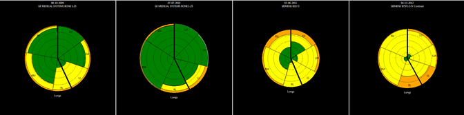
Results: We identified 172 patients with IIM and ILD (62% female; mean age 51 years; mean follow-up 4.1 years). Of those, 59 had DM, 84 had PM, 22 had anti-synthetase syndrome, and 7 were unknown. HRCT images revealed ILD in 95% of patients. Main ILD types were nonspecific interstitial pneumonia (59%), cryptogenic organizing pneumonia (23%), and usual interstitial pneumonia (9%). At year 1, improvement occurred in HRCT scans (n = 58) in percentage of reticular densities from 7 ± 6% to 5 ± 6% (p < 0.001) and interstitial abnormalities from 20 ± 16% to 17 ± 16% (p = 0.049), and a trend in improvement in DLCO (n = 80) from 56.2 ± 18.1 to 58.8 ± 16.6 percent predicted (p = 0.053). Baseline to year 3, improvement occurred (n = 34) in reticular densities (p = 0.011) but no difference in PFT values (n = 64). At year 1, 22 patients met DOI; within these, there was improvement in DLCO (n = 22): 46.8 ± 10.8 to 62.5 ± 12.8 (p <0.001) and TLC (n = 12): 65.9 ± 15.4 to 73.9 ± 13.4 (p = 0.004). Subgroup analysis by treatment, prednisone (group 1; n = 43), prednisone + other immunosuppressant (group 2; n = 65), other immunosuppressant (group 3; n = 18), and none (group 4; n = 40) revealed no differences in PFTs at baseline; but, in group 4 there were more interstitial abnormalities at baseline (p = 0.050). Baseline to year 3, group 4 worsened in interstitial abnormalities (p = 0.036). Progression from normal lung (green) to ground glass opacities (yellow) and reticular fibrosis (orange) also occurred despite treatment (See Figure).

Conclusion: IIM patients with ILD improve in reticular densities, low attenuation areas, and interstitial abnormalities. Progression of ILD was observed in patients without treatment. CALIPER aids in tracking ILD over time.

Figure. CALIPER in ILD.


Disclosure:

K. Wilton,
None;

B. Bartholmai,
None;

S. Kalra,
None;

C. S. Crowson,
None;

H. Khun,
None;

F. C. Ernste,
None.

  • Tweet
  • Click to email a link to a friend (Opens in new window) Email
  • Click to print (Opens in new window) Print

« Back to 2013 ACR/ARHP Annual Meeting

ACR Meeting Abstracts - https://acrabstracts.org/abstract/use-of-computer-aided-lung-informatics-for-pathology-evaluation-and-rating-software-in-high-resolution-computed-tomography-in-patients-with-idiopathic-inflammatory-myopathy-and-inter/

Advanced Search

Your Favorites

You can save and print a list of your favorite abstracts during your browser session by clicking the “Favorite” button at the bottom of any abstract. View your favorites »

All abstracts accepted to ACR Convergence are under media embargo once the ACR has notified presenters of their abstract’s acceptance. They may be presented at other meetings or published as manuscripts after this time but should not be discussed in non-scholarly venues or outlets. The following embargo policies are strictly enforced by the ACR.

Accepted abstracts are made available to the public online in advance of the meeting and are published in a special online supplement of our scientific journal, Arthritis & Rheumatology. Information contained in those abstracts may not be released until the abstracts appear online. In an exception to the media embargo, academic institutions, private organizations, and companies with products whose value may be influenced by information contained in an abstract may issue a press release to coincide with the availability of an ACR abstract on the ACR website. However, the ACR continues to require that information that goes beyond that contained in the abstract (e.g., discussion of the abstract done as part of editorial news coverage) is under media embargo until 10:00 AM ET on November 14, 2024. Journalists with access to embargoed information cannot release articles or editorial news coverage before this time. Editorial news coverage is considered original articles/videos developed by employed journalists to report facts, commentary, and subject matter expert quotes in a narrative form using a variety of sources (e.g., research, announcements, press releases, events, etc.).

Violation of this policy may result in the abstract being withdrawn from the meeting and other measures deemed appropriate. Authors are responsible for notifying colleagues, institutions, communications firms, and all other stakeholders related to the development or promotion of the abstract about this policy. If you have questions about the ACR abstract embargo policy, please contact ACR abstracts staff at [email protected].

Wiley

  • Online Journal
  • Privacy Policy
  • Permissions Policies
  • Cookie Preferences

© Copyright 2025 American College of Rheumatology