Session Information
Session Type: Poster Session C
Session Time: 9:00AM-11:00AM
Background/Purpose: PET/CT is a highly sensitive and specific test for diagnosing PMR.(1) This study investigated the utility of 18F-FDG whole body PET/CT in patients presenting with atypical clinical features and tested the performance of a novel artificial intelligence (AI) algorithm to simplify PMR diagnosis.
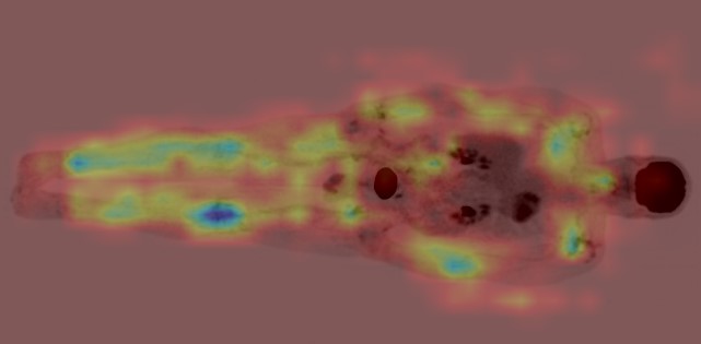
Methods: PET/CT reports from January 2015 to current were searched for findings suggestive of PMR using natural language processing. The scanned medical record of each identified case was reviewed to determine the final clinical diagnosis. Required criteria per the 2012 EULAR/ACR PMR Classification Criteria(2) (age ≥50 years, bilateral shoulder aching and abnormal CRP +/ ESR) were recorded, with patients not meeting these considered “atypical”. Qualitative and semi-quantitative (SUVmax) PET/CT scores of steroid-naïve atypical PMR patients were compared with an existing typical steroid-naïve PMR population. Differences in 18F-FDG avidity (mean SUVmax) were examined at key musculoskeletal sites. The performance of a ResNet50 AI model trained on PET/CT images of typical PMR and large joint arthritis patients was tested on the atypical PMR cohort. GradCam maps were employed to cross-check the AI’s focus on characteristic PMR-involved anatomical regions.
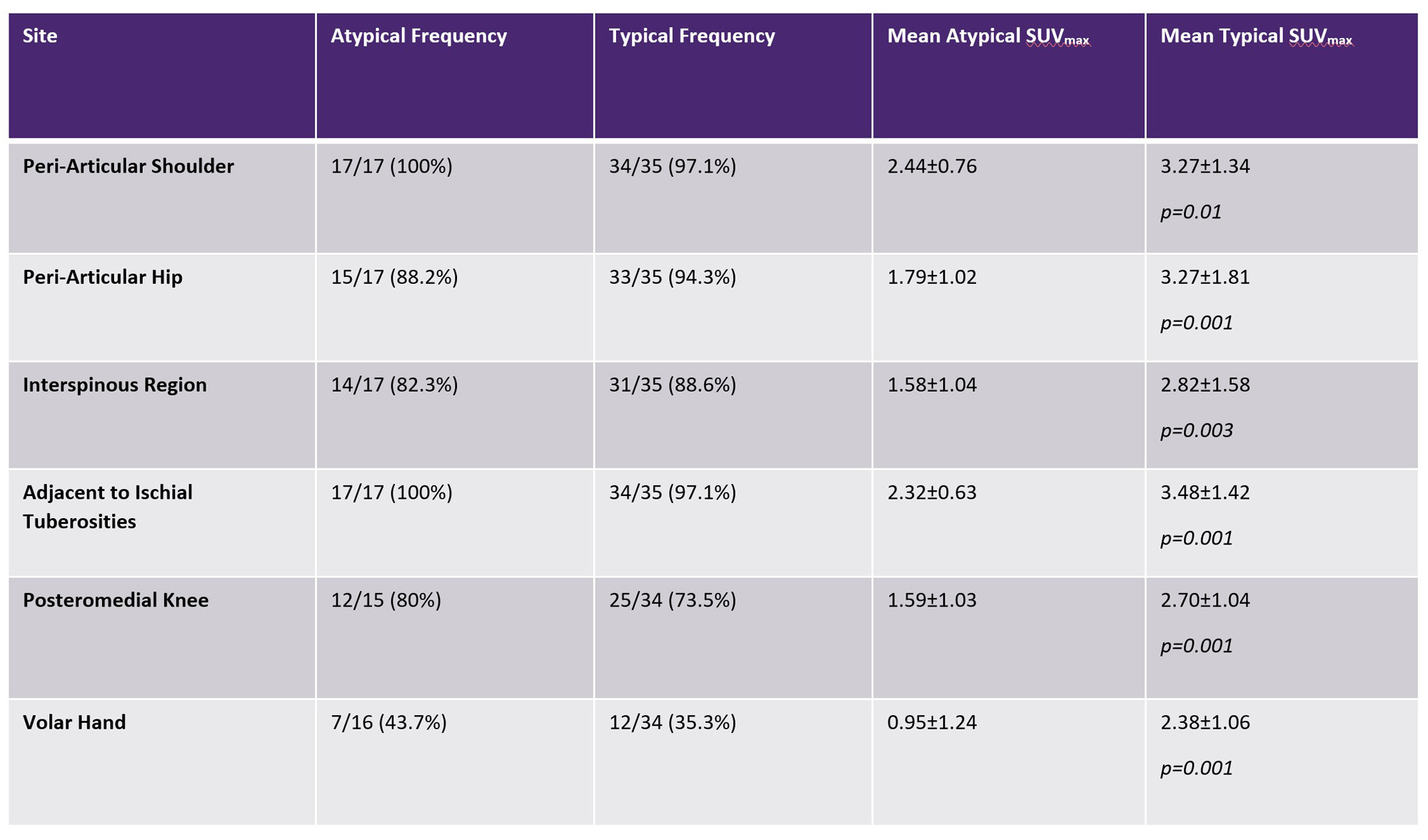
Results: 225 PET/CT reports were retrieved from >38,000 scans, with 121 patients possessing a final clinical PMR diagnosis. 17 steroid-naïve atypical patients were compared with 35 steroid-naïve typical cases. Mean CRP (10.1±20.9 cf. 44.0±32.9 [p=0.0002]) and ESR (11.5±7.2 cf. 49.3±29.6 [p=0.0001]) were lower in the atypical group, but the frequency of abnormalities at key musculoskeletal sites was similar (Table 1). A statistically significant difference (p < 0.05) in mean SUVmax was detected between the groups at every musculoskeletal site analyzed. When the novel AI-trained model was tested on PET/CT images of atypical patients, 16/17 were identified as positive for a PMR diagnosis. Figure 1 provides a GradCam map example in an atypical PMR case.
Conclusion: PMR patients with atypical clinical features exhibit the same PET/CT abnormalities as typical cases, but affected musculoskeletal sites are less 18F-FDG avid. This distinctive imaging pattern can be harnessed by AI technology to facilitate PMR diagnosis in challenging clinical scenarios.
References:
1. Owen CE, Poon AMT, Yang V, McMaster C, Lee ST, Liew DFL, et al. Abnormalities at three musculoskeletal sites on whole-body positron emission tomography/computed tomography can diagnose polymyalgia rheumatica with high sensitivity and specificity. Eur J Nucl Med Mol Imaging. 2020. 2. Dasgupta B, Cimmino MA, Kremers HM, Schmidt WA, Schirmer M, Salvarani C, et al. 2012 Provisional classification criteria for polymyalgia rheumatica: a European League Against Rheumatism/American College of Rheumatology collaborative initiative. Arthritis Rheumatol. 2012;64(4):943-54.
To cite this abstract in AMA style:
Martin E, McMaster C, Rowson S, Yang V, Poon A, Liu B, Leung J, Scott A, Liew D, Owen C. Use of Artificial Intelligence to Diagnose Polymyalgia Rheumatica on 18F-FDG Whole Body PET/CT in Patients with Atypical Clinical Features [abstract]. Arthritis Rheumatol. 2023; 75 (suppl 9). https://acrabstracts.org/abstract/use-of-artificial-intelligence-to-diagnose-polymyalgia-rheumatica-on-18f-fdg-whole-body-pet-ct-in-patients-with-atypical-clinical-features/. Accessed .« Back to ACR Convergence 2023
ACR Meeting Abstracts - https://acrabstracts.org/abstract/use-of-artificial-intelligence-to-diagnose-polymyalgia-rheumatica-on-18f-fdg-whole-body-pet-ct-in-patients-with-atypical-clinical-features/


