Session Information
Date: Monday, November 9, 2020
Title: Vasculitis – Non-ANCA-Associated & Related Disorders Poster II
Session Type: Poster Session D
Session Time: 9:00AM-11:00AM
Background/Purpose: Ultrasound (US) plays an important role in diagnosis of giant cell arteritis (GCA). To date it is unknown how intima-media-thickness (IMT) of affected arteries changes under therapy and time.
Methods: Prospective US examination of the common superficial temporal artery and the axillary artery in patients with newly diagnosed GCA, at time of diagnosis, three and six months later. All patients fulfilled ACR criteria for GCA and showed IMT values above published cut-off values of the respective artery 1. On each visit US-examination with IMT measurement of the arteries mentioned above was performed. Glucocorticoid dose and c-reactive protein (CRP) values were recorded.
Patients were divided into four subgroups: Group 1, GCA and affection of large and intermediate vessels, group 2, GCA with affection of intermediate vessels only, group 3, GCA and polymyalgia rheumatica (PMR) and affection of large and intermediate vessels and group 4, GCA and PMR and affection of intermediate vessels only.
Results: We included a total of 30 patients, 18 (60 %) females. Mean age was 74 years (SD±10). At time of diagnosis, mean CRP-values were 59.9 mg/L (SD±58.8) and mean glucocorticoid dose was 171.3 mg (SD±312.5) per day. After three months, mean CRP was 8.2 mg/L (SD±16.4) and mean glucocorticoid dose was 10.7 mg (SD±4.4) per day while after six months mean CRP was 1.8 mg/L (SD±2.1) and mean glucocorticoid dose was 3.5 mg (SD±2.9) per day.
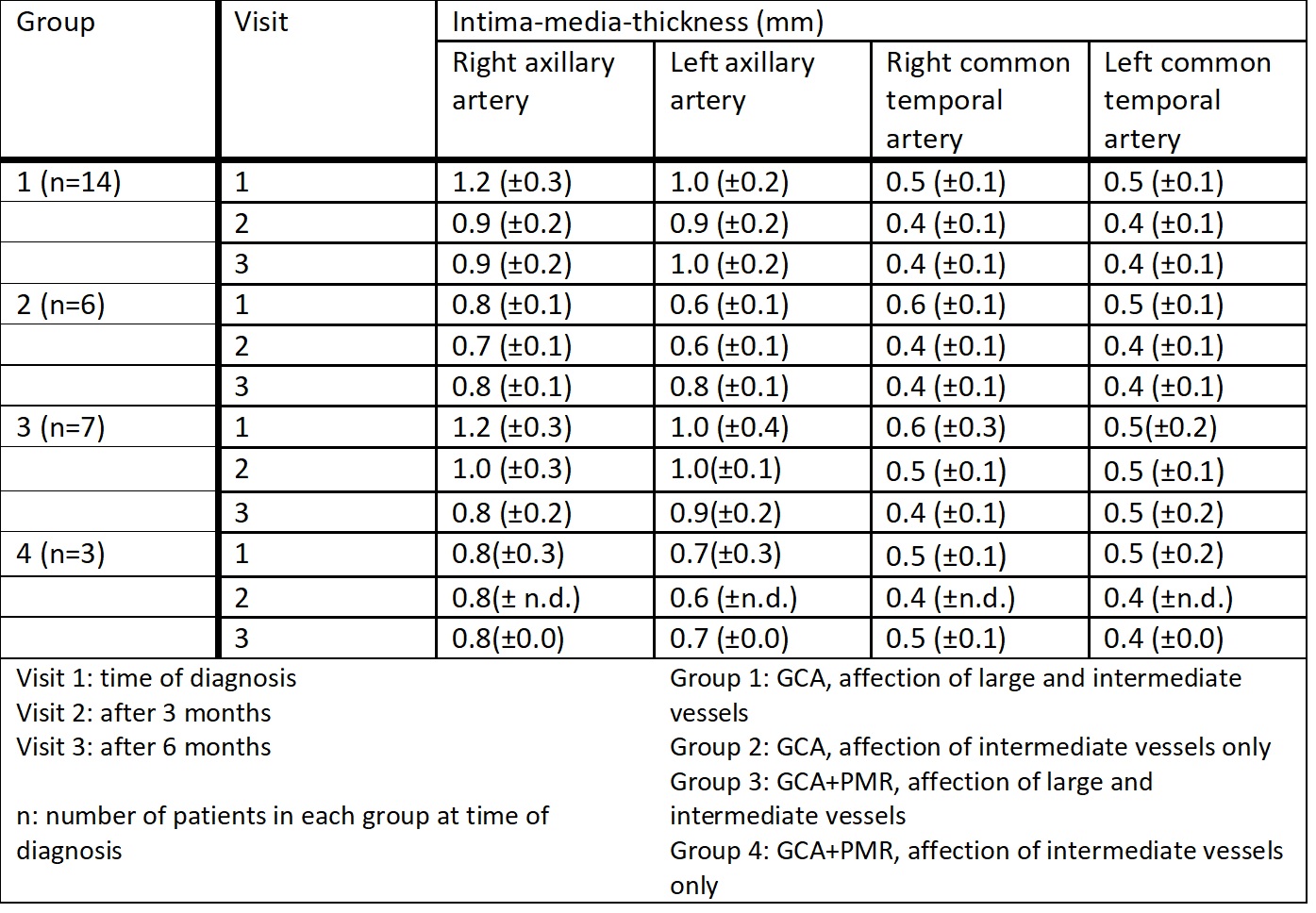
Overall a decrease of IMT values over three and six months could be observed. Exact values of each group are depicted in Table 1. Figure 1 and 2 show the decrease of IMT-values with the use of boxplots.
Conclusion: A relevant decrease of IMT-values over 6 months of therapy was observed in axillary and common superficial temporal arteries. During the first 3 months, IMT decrease was greatest. Mean CRP-values and glucocorticoid doses decreased over time and therapy. Further research in other arteries typically affected in GCA and a longer observation time in a larger cohort is obligate.
References
- Schäfer VS, Juche A, Ramiro S, Krause A, Schmidt WA. Ultrasound cut-off values for intima-media thickness of temporal, facial and axillary arteries in giant cell arteritis. Rheumatology (Oxford) 2017;56:1479–83.
 Table1: Reduction of intima-media-thickness of the axillary and common superficial temporal artery for each group over an observation time of 6 months.
 Figure 1: Boxplot showing decrease of intima-media-thickness of right and left axillary artery over 6 months between groups.
 Figure 2: Boxplot showing decrease of intima-media-thickness of right and left common superficial temporal artery over 6 months between groups.
To cite this abstract in AMA style:
Burg L, Brossart P, Behning C, Schaefer V. Ultrasound Follow-up Examination of Intima-Media-Thickness of the Temporal and Axillary Artery over Six Months in Patients with Newly Diagnosed Giant Cell Arteritis [abstract]. Arthritis Rheumatol. 2020; 72 (suppl 10). https://acrabstracts.org/abstract/ultrasound-follow-up-examination-of-intima-media-thickness-of-the-temporal-and-axillary-artery-over-six-months-in-patients-with-newly-diagnosed-giant-cell-arteritis/. Accessed .« Back to ACR Convergence 2020
ACR Meeting Abstracts - https://acrabstracts.org/abstract/ultrasound-follow-up-examination-of-intima-media-thickness-of-the-temporal-and-axillary-artery-over-six-months-in-patients-with-newly-diagnosed-giant-cell-arteritis/
