Session Information
Date: Monday, November 14, 2022
Title: Metabolic and Crystal Arthropathies – Basic and Clinical Science Poster
Session Type: Poster Session D
Session Time: 1:00PM-3:00PM
Background/Purpose: In recent years, ultrasonography (US) has emerged as an accurate and reliable tool for the diagnosis of calcium pyrophosphate (CPP) deposition disease (CPPD) in daily practice. Previous studies analyzed the diagnostic value of US findings in different tissues and joints. However, there is only scarce evidence on the US scanning protocol with the best accuracy for the diagnosis of CPPD at patient level. The aim of this study was to develop an US scanning protocol with the highest diagnostic performance for diagnosing CPPD in inter-critical periods.
Methods: Patients with a crystal-proven diagnosis of CPPD and age- and sex-matched disease-controls with a negative synovial fluid analysis for CPP crystals were prospectively enrolled in this cross-sectional, multicenter, case-control study. All subjects underwent a bilateral US examination of 9 hyaline cartilages (HC), 6 fibrocartilages (FC), 5 tendons, 1 joint recess and 1 ligament as follows: shoulder (glenoid FC, humeral HC and acromioclavicular FC), elbow (humeral HC), wrist (triangular FC, scapho-lunate ligament, volar recess of the radio-lunate joint), hand (HC of the metacarpophalangeal joints from 2nd to 5th finger), hip (acetabular FC and femoral HC), knee (femoral condyles’ HC, meniscal FC, patellar tendon), ankle (talar HC, Achilles tendon, medial and lateral compartments’ tendons, and plantar fascia). US assessment was carried-out by rheumatologists blinded to clinical and laboratory data. CPP deposits were identified as presence/absence, according to the OMERACT definitions [Filippou G, et al. Ann Rheum Dis. 2018;77:1194-9]. The diagnostic accuracy of the scanning protocols was evaluated in terms of sensitivity (SE), specificity (SP), positive and negative likelihood ratios (LH+ and LH-, respectively) and their 95% confidence intervals (95%CI). Reduced scanning protocols were defined using a stepwise backward selection. To assess the diagnostic performance of reduced scanning protocols, we compared the area under the ROC curves (AUROCs). In case of overlapping AUROCs, the shortest scanning protocol was selected.
Results: One hundred and fifty-four patients were enrolled: 88 CPPD patients (age: 70.3±12.1 years, disease duration: 4.4±5.2 years, female/male ratio: 1.7) and 66 age- and sex-matched disease-controls (age: 69.1±13.5 years, female/male ratio: 1.6).
Table 1 shows the prevalence of CPP deposits in patients with CPPD in each anatomical site included in the scanning protocol, with the FC of the medial and lateral meniscus of the knee being the most frequently involved targets (87.5% and 80.7% of patients, respectively).
Table 2 shows the diagnostic accuracy of the different US scanning protocols, with the best tradeoff between sensitivity and specificity being the identification of CPP deposits in at least two anatomical sites both in the entire protocol (SE 96.6% [95%CI 90.4-99.3%]; SP 92.7% [95%CI 83.7-97.6%]) and in a reduced protocol of three joints (knees, wrists and hips) (SE 93.2% [95%CI 85.8-97.5%]; SP 98.5% [95%CI 92.1-99.9%]).
Conclusion: Our results show that bilateral US assessment of knees, wrists and hips provided the best sensitivity and specificity for diagnosing CPPD.

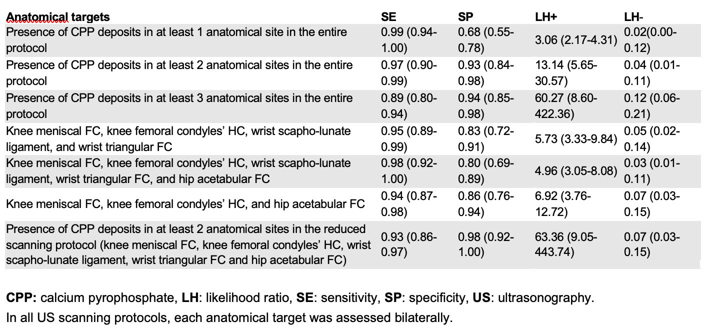
In all US scanning protocols, each anatomical target was assessed bilaterally.
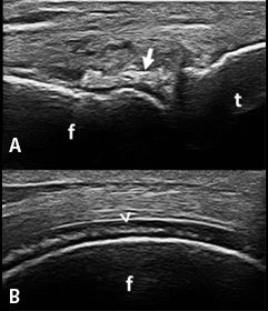
A: Knee, longitudinal scan of the lateral meniscus.
B: Knee, longitudinal scan of the medial femoral condyle’s HC.
Arrows: CPP crystal deposits at FC, arrowhead: CPP crystal deposits at HC.
To cite this abstract in AMA style:
Cipolletta E, Moscioni E, Sirotti S, Filippou G, Filippucci E. Ultrasound Diagnosis of Calcium Pyrophosphate Crystal Deposition: Which Sites Should Be Scanned? [abstract]. Arthritis Rheumatol. 2022; 74 (suppl 9). https://acrabstracts.org/abstract/ultrasound-diagnosis-of-calcium-pyrophosphate-crystal-deposition-which-sites-should-be-scanned/. Accessed .« Back to ACR Convergence 2022
ACR Meeting Abstracts - https://acrabstracts.org/abstract/ultrasound-diagnosis-of-calcium-pyrophosphate-crystal-deposition-which-sites-should-be-scanned/