Session Information
Date: Tuesday, November 12, 2019
Title: Vasculitis – Non-ANCA-Associated & Related Disorders Poster III: Giant Cell Arteritis
Session Type: Poster Session (Tuesday)
Session Time: 9:00AM-11:00AM
Background/Purpose: Imaging has been recently recognized as a tool equivalent to the temporal artery biopsy in diagnosing giant cell arteritis (GCA). Amongst a variety of imaging modalities, color Doppler Ultrasonography (CDS) is the most convenient. We aimed to evaluate the frequency of cranial- and aortic arch-artery involvement in GCA using CDS.
Methods: We performed CDS examination of cranial and aortic arch arteries in new, clinically diagnosed GCA patients between October 2013 and April 2019, using a Philips IU22 with 5–17.5 MHz linear probe or Philips Epiq 7 with 5–18.5 MHz linear probe. Temporal, facial, occipital, thyroid, carotid, vertebral, subclavian, and axillary arteries were examined bilaterally. A halo with positive compression sign was considered a positive finding. Additionally, the thickness of intima-media complex was measured. .
Results: During the 67-month period 212 newly diagnosed GCA patients (63.2% females, median (IQR) age 75.4 (67.2-80.8) years) underwent vascular CDS evaluation. CDS was performed prior to glucocorticoid initiation in 201 patients (94.8%), and delayed by a median (IQR) of 1 (1-3) days (range 1-6 days) for the rest.
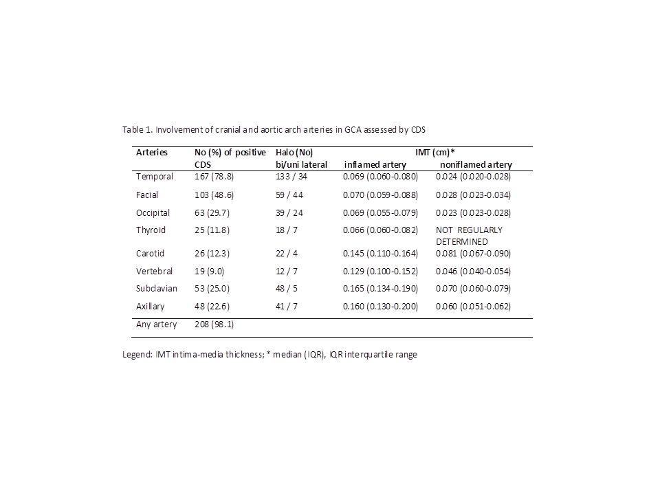
The CDS was positive in 208 (98.1%) patients in at least one of the examined arteries. Temporal arteries, involved in 167 (78.8%) patients, were the most commonly affected vessels. Extracranial large vessel involvement (LVV) was found in 70 (33.0%) patients (30 patients had isolated LVV, and 40 concomitant temporal artery involvement). Among the 142 patients without LVV, 11(5.2% of the studied cohort) had involvement of cranial arteries other than temporal arteries (we found facial, occipital and thyroid artery involvement in 9, 3 and 2 patients, respectively). Table 1 shows the frequency of individual vessel involvement and the intima-media thickness of inflamed and non-inflamed arteries.
Conclusion: CDS of eight preselected cranial and aortic arch arteries provides a high diagnostic yield in GCA.
To cite this abstract in AMA style:
JESE R, Rotar Z, Tomšič M, Hocevar A. Ultrasonography in the Diagnosis of Giant Cell Arteritis [abstract]. Arthritis Rheumatol. 2019; 71 (suppl 10). https://acrabstracts.org/abstract/ultrasonography-in-the-diagnosis-of-giant-cell-arteritis/. Accessed .« Back to 2019 ACR/ARP Annual Meeting
ACR Meeting Abstracts - https://acrabstracts.org/abstract/ultrasonography-in-the-diagnosis-of-giant-cell-arteritis/

