Session Information
Session Time: 6:00PM-7:00PM
Background/Purpose: Juvenile idiopathic arthritis (JIA) is the most common pediatric rheumatologic disease, with an incidence of 2–23 new cases per 100,000 children, and is associated with significant morbidity. Analyses of peripheral blood and synovial fluid have provided limited insight into JIA pathogenesis and have yet to identify reliable biomarkers to predict disease course and/or drug response. Novel approaches utilizing minimally invasive ultrasound-guided synovial biopsy (MIUGSB) allow direct analysis of the synovial tissue, which is the primary site of inflammation in JIA.
Methods: Patients with JIA, who were at least 5 years old and were undergoing ultrasound-guided intra-articular corticosteroid injection (IACI), were eligible for synovial tissue biopsy. This study was approved by Lurie Children’s Hospital IRB. At time of IACI, five to eight samples of synovial tissue from a single knee per patient were retrieved via MIUGSB by a pediatric rheumatologist. Synovial tissue samples were either formalin-fixed and paraffin-embedded (FFPE) for spatial transcriptomic profiling on the 10x Genomics Xenium platform using a custom 216-gene panel, or enzymatically digested and sorted for generation of single cell 5’ libraries and RNA sequencing.
Results: We obtained high quality knee synovial biopsies from 17 patients with oligoarticular or polyarticular JIA (Table 1); all patients tolerated the procedure well. Single-cell RNA sequencing (scRNA-seq) detected a median of 1,054 to 3,632 genes per cell from 10 patients, with data currently under analysis. To date, spatial transcriptomic analysis has been performed on synovial tissue from 4 of these patients. In addition, FFPE synovial tissue samples from 5 additional patients have been prepared for Xenium-based spatial transcriptomics. We identified 11 clusters that comprise diverse immune cell populations—including monocytes, macrophages, T-, B-, plasma, and dendritic cells—organized regionally in relation to the synovial lining and sublining layers. In contrast to samples from patients with rheumatoid arthritis (RA) in which fibroblast-like synoviocytes outnumber macrophages, most synovial cells from JIA samples were macrophages (Figure 1). Future studies will delineate the topography of the individual subpopulations in JIA synovium compared to RA synovium.
Conclusion: This study demonstrates the feasibility of concomitantly leveraging scRNA-seq to identify transcriptional signatures and spatial transcriptomics to characterize immune-stromal architectures associated with JIA disease. Additionally, these findings will be correlated with prospective clinical data as well as analysis from peripheral blood and synovial fluid, offering insight into pediatric synovial immunopathology and potential insight into predictors of immune response.
Table 1. Clinical Characteristics of Patient Participants (n=17)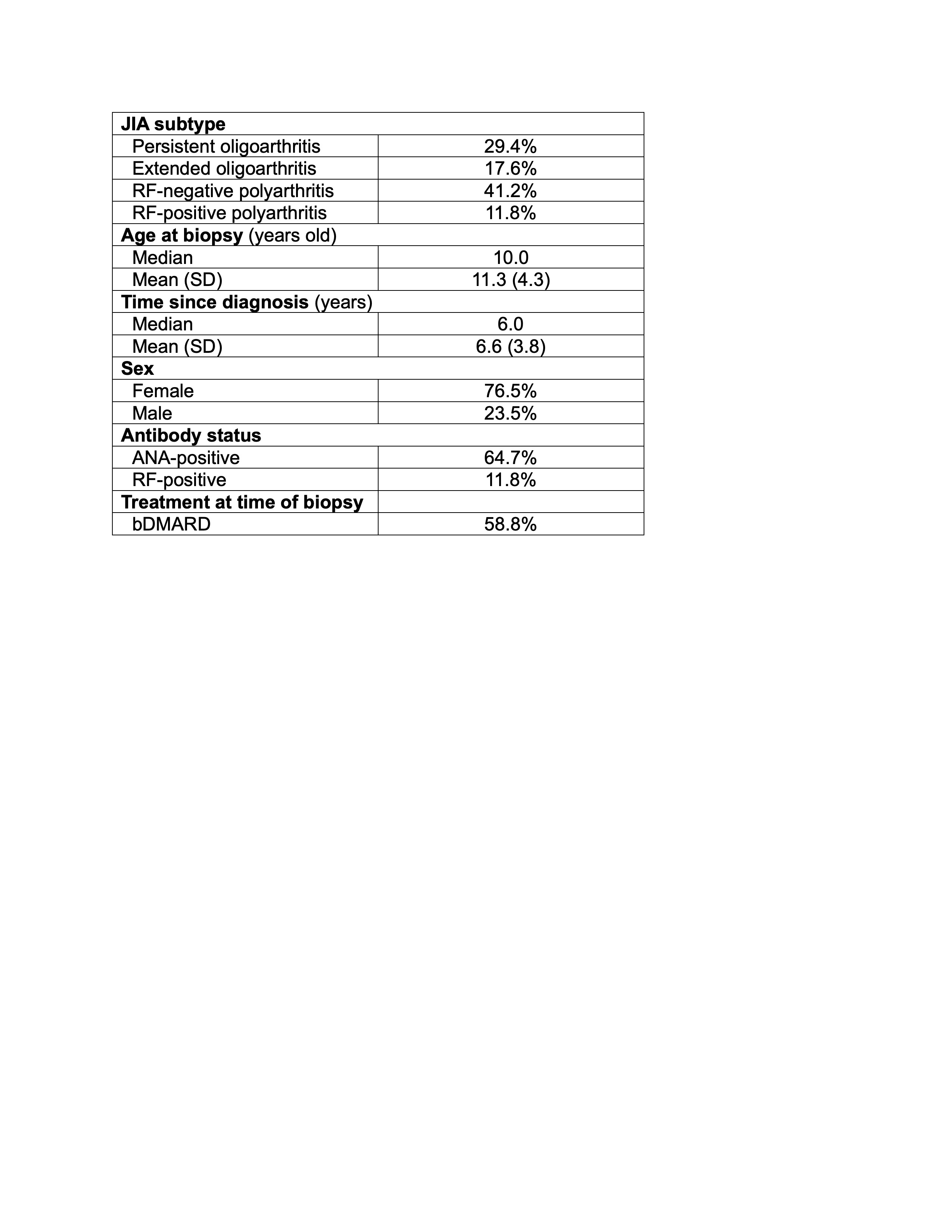
Figure 1. Characterization of Immune and Non-immune cells from Knee Synovium of Patients with JIA A, Histology of synovial tissue, illustrating representative hematoxylin and eosin (H&E), anti-CD45 immunohistochemistry (IHC), and anti-CD68 IHC. B, Uniform Manifold Approximation and Projection (UMAP) of annotated cell types from reference CosMx dataset for label transfer to Xenium dataset. C, Spatial plot of JIA synovial tissue, illustrating representative cell annotation.
To cite this abstract in AMA style:
Dowling S, Khan M, Therron T, Dapas M, Nafikova R, Mian K, DeRanieri D, Winter D, Perlman H. Transcriptomic Analysis of Ultrasound-Guided Synovial Biopsies from Patients with Juvenile Idiopathic Arthritis [abstract]. Arthritis Rheumatol. 2026; 78 (suppl 3). https://acrabstracts.org/abstract/transcriptomic-analysis-of-ultrasound-guided-synovial-biopsies-from-patients-with-juvenile-idiopathic-arthritis/. Accessed .« Back to 2026 Pediatric Rheumatology Symposium
ACR Meeting Abstracts - https://acrabstracts.org/abstract/transcriptomic-analysis-of-ultrasound-guided-synovial-biopsies-from-patients-with-juvenile-idiopathic-arthritis/