Session Information
Session Type: Poster Session C
Session Time: 10:30AM-12:30PM
Background/Purpose: Chronic nonbacterial osteomyelitis (CNO) of the clavicle can pose a diagnostic challenge as the differential includes malignancy and infection. Biopsy is often required for unifocal clavicular lesions as CNO is a diagnosis of exclusion. With recent advances in imaging techniques, we aim to describe features on plain radiograph and MRI that may distinguish CNO from other conditions that must be excluded.
Methods: This is a single-centre retrospective chart review of all patients presenting with a unifocal clavicular lesion who underwent a clavicular biopsy and radiographic imaging between the years of 2000-2022. A diagnosis of CNO or non-CNO was extracted from chart review based on histology and the opinion of the treating physician. Imaging (plain radiograph, MRI) was reviewed by two musculoskeletal radiologists blinded to the final diagnosis. Clinical and imaging features were compared between patients diagnosed with CNO and non-CNO diagnoses using Fishers’ Exact Test.
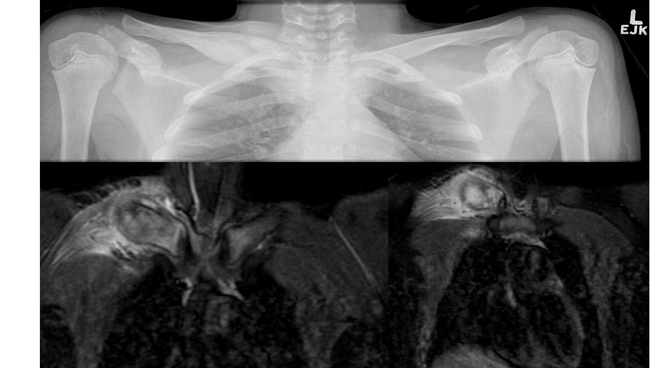
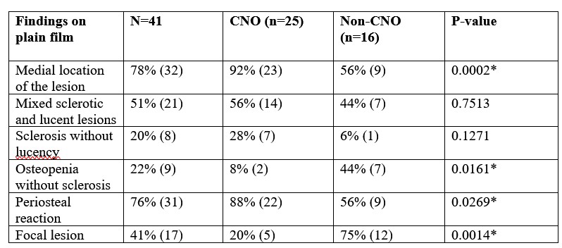
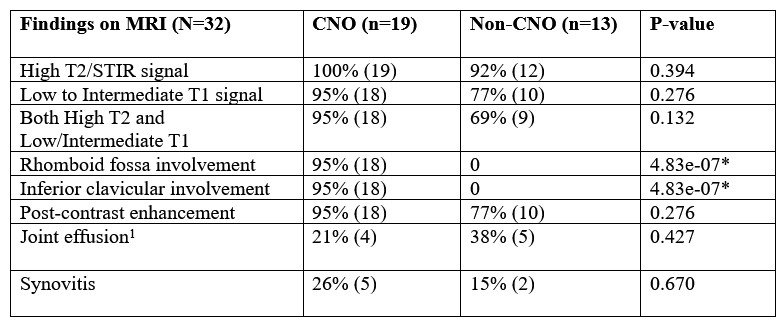
Results: 41 patients were included in the analysis: 25 patients with CNO and 16 with non-CNO (diagnoses included: aneurysmal bone cyst (n=5), Langerhans Cell Histiocytosis (n=4), leukemia (n=1), infectious osteomyelitis (n=1), Gorham Stout (n=2), fracture (n=1), fibrous dysplasia (n=1) and 1 diagnosis was unknown). All patients had radiography of the clavicles and 32 patients had MRI. Patients with CNO were more likely to have multifocal lesions on imaging (p=0.01) and were less likely to have fever or weight loss at presentation (p=0.05). CNO lesions on plain film (Fig. 1) were more likely to be located on the medial clavicle (p=2e-04) with periosteal reaction (p=0.03) (Table 2). On MRI, rhomboid fossa involvement (Fig. 1) was found in 19 of 25 patients with CNO, and 0 of 16 in those with non-CNO (p=4.87e-07) (Table 3).
Conclusion: This study describes a novel association of rhomboid fossa involvement on MRI with CNO that is not present in non-CNO cases. Multifocal lesions on imaging, absence of fever/weight loss, periosteal reaction on plain film and medial clavicular involvement are supportive features of CNO. MRI is an important diagnostic tool and it may be possible to avoid biopsy by using imaging modalities in conjunction with clinical features to make the diagnosis of CNO.
Top: Plain film showing cortical and periosteal hyperostosis in the medial right clavicle. Associated mild periosteal reaction with bony expansion.
Bottom: Coronal STIR images demonstrating increased bone marrow signal with extensive periosteal reaction and cortical thickening in the medial right clavicle. Associated soft tissue edema in the surrounding muscles with increased signal in the right rhomboid fossa.
To cite this abstract in AMA style:
Chen A, Hameed S, Hadi A, Hopyan S, Somers G, Laxer R, Stimec J. To Biopsy or Not to Biopsy: Imaging Features of Chronic Nonbacterial Osteomyelitis of the Clavicle [abstract]. Arthritis Rheumatol. 2024; 76 (suppl 9). https://acrabstracts.org/abstract/to-biopsy-or-not-to-biopsy-imaging-features-of-chronic-nonbacterial-osteomyelitis-of-the-clavicle/. Accessed .« Back to ACR Convergence 2024
ACR Meeting Abstracts - https://acrabstracts.org/abstract/to-biopsy-or-not-to-biopsy-imaging-features-of-chronic-nonbacterial-osteomyelitis-of-the-clavicle/


