ACR Meeting Abstracts

ACR Meeting Abstracts

  • Meetings
    • ACR Convergence 2024
    • ACR Convergence 2023
    • 2023 ACR/ARP PRSYM
    • ACR Convergence 2022
    • ACR Convergence 2021
    • ACR Convergence 2020
    • 2020 ACR/ARP PRSYM
    • 2019 ACR/ARP Annual Meeting
    • 2018-2009 Meetings
    • Download Abstracts
  • Keyword Index
  • Advanced Search
  • Your Favorites
    • Favorites
    • Login
    • View and print all favorites
    • Clear all your favorites
  • ACR Meetings

Abstract Number: 809

TNF-α Drives Progressive Obliterative Pulmonary Vascular Disease and Represents a Novel Model of Connective-Tissue Disease Associated Pulmonary Arterial Hypertension (CTD-PAH)

Benjamin Korman1, Richard Bell 1, R James White 2, Maria de la luz Garcia-Hernandez 3, Emily Wu 2, Pamela Slattery 2, Nelson Huertas 2, Stacey Duemmel 2, Marc Nuzzo 2, Homaira Rahimi 4, Craig Morrell 2, Christopher Ritchlin 3 and Edward Schwarz 2, 1University of Rochester Medical Center, Rochester, NY, 2University of Rochester, Rochester, NY, 3Division of Allergy, Immunology and Rheumatology, Center for Musculoskeletal Research, University of Rochester School of Medicine and Dentistry, Rochester, NY, USA, Rochester, NY, 4University of Rochester Medical Center, Rochester

Meeting: 2019 ACR/ARP Annual Meeting

Keywords: Connective tissue diseases, pulmonary complications, scleroderma-like conditions, tumor necrosis factor (TNF) and mouse model

  • Tweet
  • Email
  • Print
Session Information

Date: Sunday, November 10, 2019

Title: 3S032: Plenary I (805–809)

Session Type: Plenary Session I

Session Time: 11:00AM-12:30PM

Background/Purpose: Pulmonary arterial hypertension (PAH) is a severe cardiopulmonary disease characterized by an obliterative vasculopathy and vascular remodeling, right heart hypertrophy, and premature death.  Connective tissue disease associated PAH (CTD-PAH) is common in scleroderma, and other autoimmune diseases, which have poor clinical outcomes.   Recently, it has been shown that female human TNF-transgenic (TNF-Tg) mice die by 6-months from cardiopulmonary disease.  Thus, we aimed to formally characterize this pathophysiology and assess its potential as a model of CTD-PAH.

Methods: Histologic analysis and immunofluorescent (IF) staining was performed on female TNF-Tg (3647 line) and wild type (WT) mice to characterize the pulmonary vascular and right ventricular pathology.  Mice (n > 4) underwent: right heart catheterization to assess hemodynamics, or barium-perfused micro-CT to assess vascular morphology, or gas chromatography.  Lungs from TNF-Tg/WT bone marrow chimeric mice, and anti-TNF vs. placebo treated TNF-Tg mice were assessed (n > 3).  RNA sequencing was performed on lung tissue, and bioinformatic techniques were applied to compare TNF-Tg mouse lungs to publicly available human normal and CTD-PAH transcriptomic data.

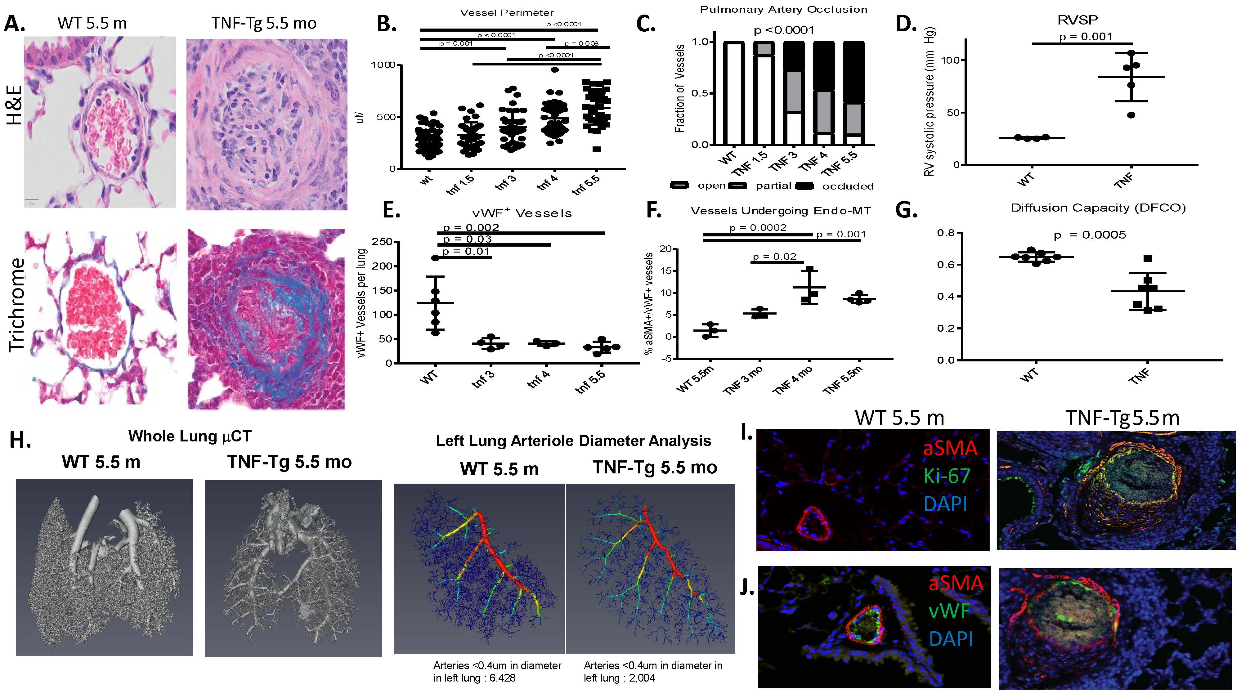
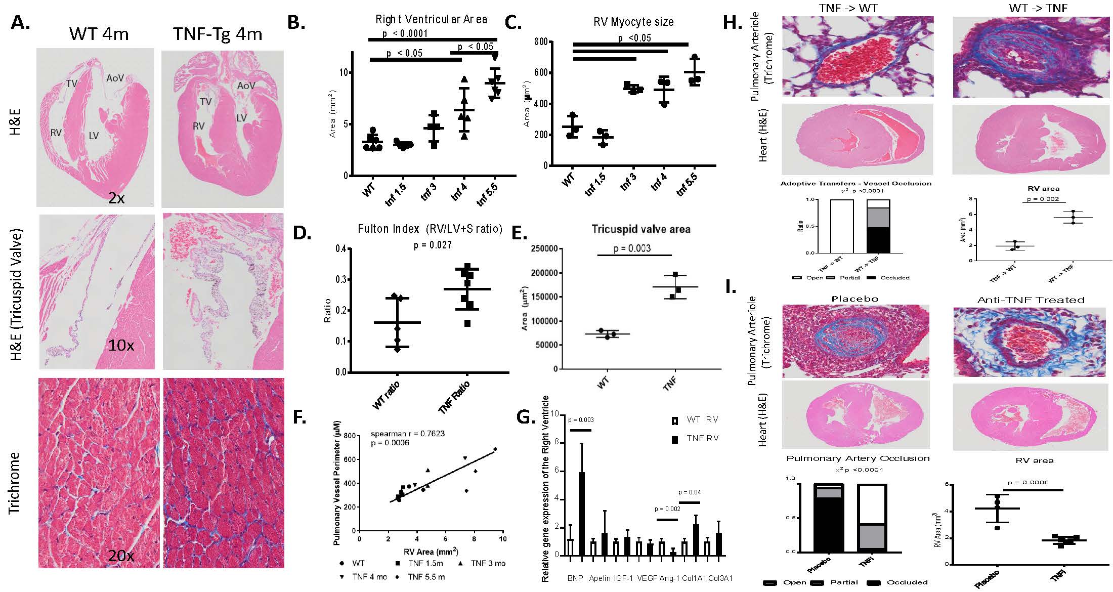
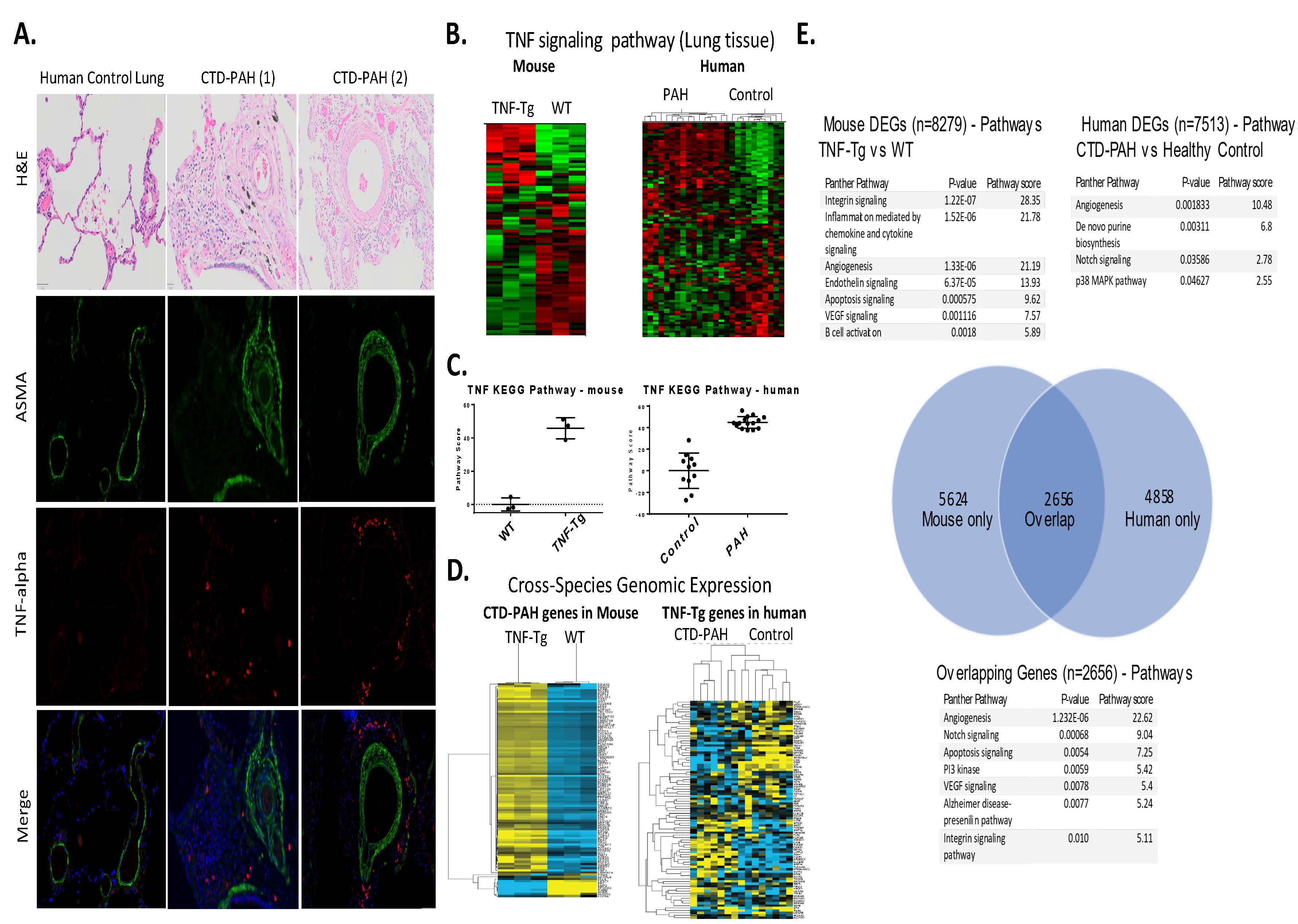
Results: Female TNF-Tg mice display a progressive pulmonary vasculopathy beginning at 3 months of age manifested by vascular collagen deposition, enlarged pulmonary arteries, attenuation of distal arterioles, and vascular occlusion, which closely resemble CTD-PAH pathologically (Fig 1A-C).  Hemodynamic assessment demonstrated a significantly increased right ventricular systolic pressure of 83.7± 10.3 mmHg vs. 25.7 ± 0.4 mmHg in TNF-Tg vs. WT mice (Fig 1D), making this one of the most robust models of murine PAH ever reported. μCT analysis confirmed pruning of the vascular tree (Fig 1H), and TNF-Tg mice had reduced gas exchange (Fig 1G).  Increased aSMA IF staining in TNF-Tg lungs corresponded to proliferation (Ki-67+), and loss of von Willebrand factor positive (vWF+) vessels over time.  We also observed an increase in aSMA+vWF+ cells (Fig 1, E, F, I), implicating endothelial-mesenchymal transition in this process.  By 4 months of age, TNF-Tg mice display remarkable right ventricular hypertrophy, and  transcriptional evidence of RV dysregulation  (Fig 2A-G).  Bone marrow chimera experiments revealed that mesenchymal cells, and not bone-marrow derived cells, are necessary to drive this process (Fig 2H), while anti-TNF therapy halted the progression of PAH pathology (Fig 2I).  Human SSc-PAH lungs display increased TNF-a staining (Fig 3A), and human microarray data demonstrated a prominent TNF signature that can distinguish PAH from control lungs (Fig 3B).  Comparison of gene expression between TNF-Tg lungs and CTD-PAH lungs showed significant similarities in expression patterns and clustering (Fig 3C) with enrichment in pathway overlaps including angiogenesis, Notch signaling, apoptosis, and VEGF signaling (Fig 3D).

Conclusion: The TNF-Tg mouse represents a novel model of CTD-PAH which recapitulates nearly all key features of the disease and can serve as a valuable tool to better study and test potential CTD-PAH therapeutics.


pah acr abstract figures korman et al 1

Figure 1. Characterization of pulmonary vascular abnormalities in TNF-transgenic -TNF-Tg- mice. A. Histologic evaluation of TNF-Tg pulmonary arterioles revealed intimal proliferation and vascular occlusion associated with vascular collagen deposition -Top, H&E, Bottom, Masson’s Trichrome, 20x-. B. Pulmonary arteriole size begins to increase at 3 months of age and becomes progressively larger over time. WT indicates 5.5 month old WT mice for this and all subsequent figures. C. Pulmonary arteries were progressively occluded in the TNF-Tg mice with over 90% of vessels showing at least partial occlusion by 4 months of age. D. Right heart catheterization of TNF-Tg mice demonstrate evidence of severe pulmonary arterial hypertension with increased right ventricular systolic pressure -RVSP- of 83.7± 10.3 mmHg vs. 25.7 ± 0.4 mmHg in TNF-Tg vs. WT mice. E. Number of arterioles in each mouse lung was counted on sections stained for von Willebrand factor -vWF- and there was substantial vessel loss by 3 months of age. F. Quantification of vessels immunofluorescently labeled double positive for both vWF and aSMA indicated that there is a progressive increase in endothelial-mesenchymal transition in TNF-Tg mice. G. TNF-Tg mice have impaired gas exchange as shown by gas chromatography for diffusion of carbon monoxide -DFCO-. H. 3D reconstruction of micro-CT scans of barium perfused TNF-Tg and WT lungs demonstrates substantial pruning of the vascular tree with loss of distal arterioles in the TNF-Tg mouse -left, whole lung reconstruction, right, tree structure diagram used for quantification of arteriole size in left lung-. I. Double immunostaining for aSMA and proliferation marker Ki-67 demonstrate co-localization in TNF-Tg mice indicating smooth cell proliferation not seen in WT mice. J. Double immunostaining for aSMA and vWF demonstrates co-localization of a subset of cells indicative of the presence of endo-MT in these lesions.


pah acr abstract figures korman et al 2

Figure 2. Right ventricular dysfunction in TNF-Tg mice. A. Right ventricular hypertrophy -top, H&E, 2x-, tricuspid valve enlargement -middle, H&E, 10x- and myocyte hypertrophy and collagen deposition -bottom, Masson’s trichrome, 20x- seen in 4 mo old TNF-Tg hearts compared to WT. B. Histomorphometry revealed progressive increase in RV size over time. C. Histomorphometry revealed progressive increase in RV myocyte size over time. D. Assessment of cardiac weight as measured by the Fulton index -RV/RV+LV+septum- demonstrates abnormal RV weight indicative of RV pathology in 4 mo old TNF-Tg and WT mice. E. Increase in tricuspid valve area in 4 month old TNF-Tg mice. F. RV area is significantly positively associated with pulmonary artery size. G. qPCR of RVs for genes previously shown to be abnormal in RV dysfunction demonstrate significant increases in B natriuretic peptide -BNP- and collagen 1A1 and decrease in angiopoietin-1 in TNF-Tg mice compared to WT, all genes relative expression were normalized to GAPDH. H. Bone marrow chimera experiments revealed that WT mice given TNF-Tg bone marrow at 6 weeks of age did not develop pulmonary or cardiac evidence of PAH at 5 months of age while TNF-Tg mice given WT bone marrow still developed the pathology indicating that mesenchymal rather than bone-marrow derived cells are responsible for the pathology. I. Treatment of TNF-Tg mice with anti-TNF therapy or placebo -CNTO12 and CNTO151, respectively, 10 mg/kg given weekly intraperitoneally from 3 months to 4.5 months of age- demonstrated a lack of progression of pulmonary vascular lesions and no significant right heart pathology after treatment with anti-TNF therapy while placebo animals developed severe PAH.


pah acr abstract figures korman et al 3

Figure 3. TNF expression and other relevant pathways overlap in human CTD-PAH and TNF-Tg mice. A. Immunostaining for aSMA and TNF-alpha in lung biopsies from CTD-PAH patients -n=2, representative of n=5- and normal lung -n=1 representative of n=2- demonstrate that CTD-PAH patients have a significant TNF+ infiltrate in the area surrounding vessels with aSMA proliferation not seen in control individuals. B. RNA sequencing of mouse lungs reveals that the TNF KEGG pathway can distinguish TNF-Tg and WT mice -n=3 per group- by hierarchical clustering -left heatmap, red = increased expression, green = decreased expression- and that the same TNF KEGG pathway in human lungs was able to distinguish patients with PAH of multiple etiologies -n=15- from control lungs -n=11- in publicly available microarray data -GSE113439, right heatmap-, C. Condensed TNF pathway scores show significant differences between cases and controls -left, mouse, right, human-. D. Top 100 differentially expressed genes from mouse and human PAH lungs are sufficient to distinguish PAH across species -left, top 100 CTD-PAH genes in mouse lung samples, right, top 100 TNF-Tg genes in human lung samples-. E. Pathway analysis of human and mouse PAH show substantial overlaps. Over one third -n = 2656- of genes differentially expressed -p<0.05 in one species- showed overlap between species. Top pathways in overlapping genes included angiogenesis, notch signaling, apoptosis, and PI3 kinase signaling.


Disclosure: B. Korman, None; R. Bell, None; R. White, None; M. Garcia-Hernandez, None; E. Wu, None; P. Slattery, None; N. Huertas, None; S. Duemmel, None; M. Nuzzo, None; H. Rahimi, None; C. Morrell, None; C. Ritchlin, AbbVie, 2, 5, 9, Amgen, 2, 5, BMS, 5, Janssen, 5, Janssen Research & Development, LLC, 2, Lilly, 5, Novartis, 5, Pfizer, 2, Pfizer Inc, 5, UCB, 2, 5; E. Schwarz, None.

To cite this abstract in AMA style:

Korman B, Bell R, White R, Garcia-Hernandez M, Wu E, Slattery P, Huertas N, Duemmel S, Nuzzo M, Rahimi H, Morrell C, Ritchlin C, Schwarz E. TNF-α Drives Progressive Obliterative Pulmonary Vascular Disease and Represents a Novel Model of Connective-Tissue Disease Associated Pulmonary Arterial Hypertension (CTD-PAH) [abstract]. Arthritis Rheumatol. 2019; 71 (suppl 10). https://acrabstracts.org/abstract/tnf-%ce%b1-drives-progressive-obliterative-pulmonary-vascular-disease-and-represents-a-novel-model-of-connective-tissue-disease-associated-pulmonary-arterial-hypertension-ctd-pah/. Accessed .
  • Tweet
  • Email
  • Print

« Back to 2019 ACR/ARP Annual Meeting

ACR Meeting Abstracts - https://acrabstracts.org/abstract/tnf-%ce%b1-drives-progressive-obliterative-pulmonary-vascular-disease-and-represents-a-novel-model-of-connective-tissue-disease-associated-pulmonary-arterial-hypertension-ctd-pah/

Advanced Search

Your Favorites

You can save and print a list of your favorite abstracts during your browser session by clicking the “Favorite” button at the bottom of any abstract. View your favorites »

All abstracts accepted to ACR Convergence are under media embargo once the ACR has notified presenters of their abstract’s acceptance. They may be presented at other meetings or published as manuscripts after this time but should not be discussed in non-scholarly venues or outlets. The following embargo policies are strictly enforced by the ACR.

Accepted abstracts are made available to the public online in advance of the meeting and are published in a special online supplement of our scientific journal, Arthritis & Rheumatology. Information contained in those abstracts may not be released until the abstracts appear online. In an exception to the media embargo, academic institutions, private organizations, and companies with products whose value may be influenced by information contained in an abstract may issue a press release to coincide with the availability of an ACR abstract on the ACR website. However, the ACR continues to require that information that goes beyond that contained in the abstract (e.g., discussion of the abstract done as part of editorial news coverage) is under media embargo until 10:00 AM ET on November 14, 2024. Journalists with access to embargoed information cannot release articles or editorial news coverage before this time. Editorial news coverage is considered original articles/videos developed by employed journalists to report facts, commentary, and subject matter expert quotes in a narrative form using a variety of sources (e.g., research, announcements, press releases, events, etc.).

Violation of this policy may result in the abstract being withdrawn from the meeting and other measures deemed appropriate. Authors are responsible for notifying colleagues, institutions, communications firms, and all other stakeholders related to the development or promotion of the abstract about this policy. If you have questions about the ACR abstract embargo policy, please contact ACR abstracts staff at [email protected].

Wiley

  • Online Journal
  • Privacy Policy
  • Permissions Policies
  • Cookie Preferences

© Copyright 2025 American College of Rheumatology