Session Information
Session Time: 6:00PM-7:00PM
Background/Purpose: Localized scleroderma (LS) is a disease of inflammatory fibrosis affecting skin and deeper tissue, but the mechanisms driving fibrosis are poorly understood. Although fibroblasts actively produce fibrotic material, disease tissues show significant immune cell infiltration with a clear role for interactions between T cells, macrophages, and fibroblasts seen in molecular data. Single cell transcriptomic skin and blood cytokine data suggests a role for interferon gamma (IFNγ) as a driver of inflammation affected through CXCL9, CXCL10, and their receptor CXCR3 as well as a role for secreted frizzled proteins (SFRP2, SFRP4) and serine protease (PRSS23), with increased transcription in fibroblast subpopulations of affected skin. The localization of these effectors in skin and their roles in initiating or propagating fibrosis is unclear. We hypothesized that these cell populations and localization of secreted proteins will show cell interactions affected by these inflammatory and fibrosis-driving signaling pathways.
Methods: Multiplex immunofluorescence (IF) assays were used to stain 4mm skin punch biopsy tissues including 21 LS (8 clinically active, 13 clinically inactive) and 12 healthy controls. Staining was performed in 2 panels with DAPI, CD3, CD68, and αSMA on both panels, CXCL9, CXCL10, and CXCR3 on panel 1 and SFRP2, SFRP4, and PRSS23 on panel 2. Cell and IF signal detection was performed by QuPath.
Results: Active localized scleroderma patient tissues show broader increased expression of SFRP4 and more specific SFRP2 and SFRP4 expression in periadnexal and perivascular regions that colocalized with CXCL9, CXCL10, and CXCR3. Clinically inactive disease tissues also showed increased SFRP4 and SFRP2 in periadnexal and perivascular regions with reduced CXCL9, CXCL10, and CXCR3, while similar regions in healthy control tissues showed low expression of each of these proteins in periadnexal and perivascular regions. Regions with increased IFN and secreted frizzled proteins are similarly enriched for T cells (CD3) and macrophages (CD68).
Conclusion: Expression of key fibroblast (secreted frizzled proteins 2/4) and inflammatory (CXCL9/10, CXCR3) biomarkers show increased expression and localization to periadnexal and perivascular niches in juvenile localized scleroderma tissues with colocalization of T cells and macrophages in clinically active patients with more focused and reduced expression and localization in inactive and healthy control tissues.
Whole Tissue Expression of SFRP4, SFRP2, PRSS23, CXCL9, CXCL10, CXCR3, aSMA, CD3, CD68 in Localized Scleroderma and Healthy Control Skin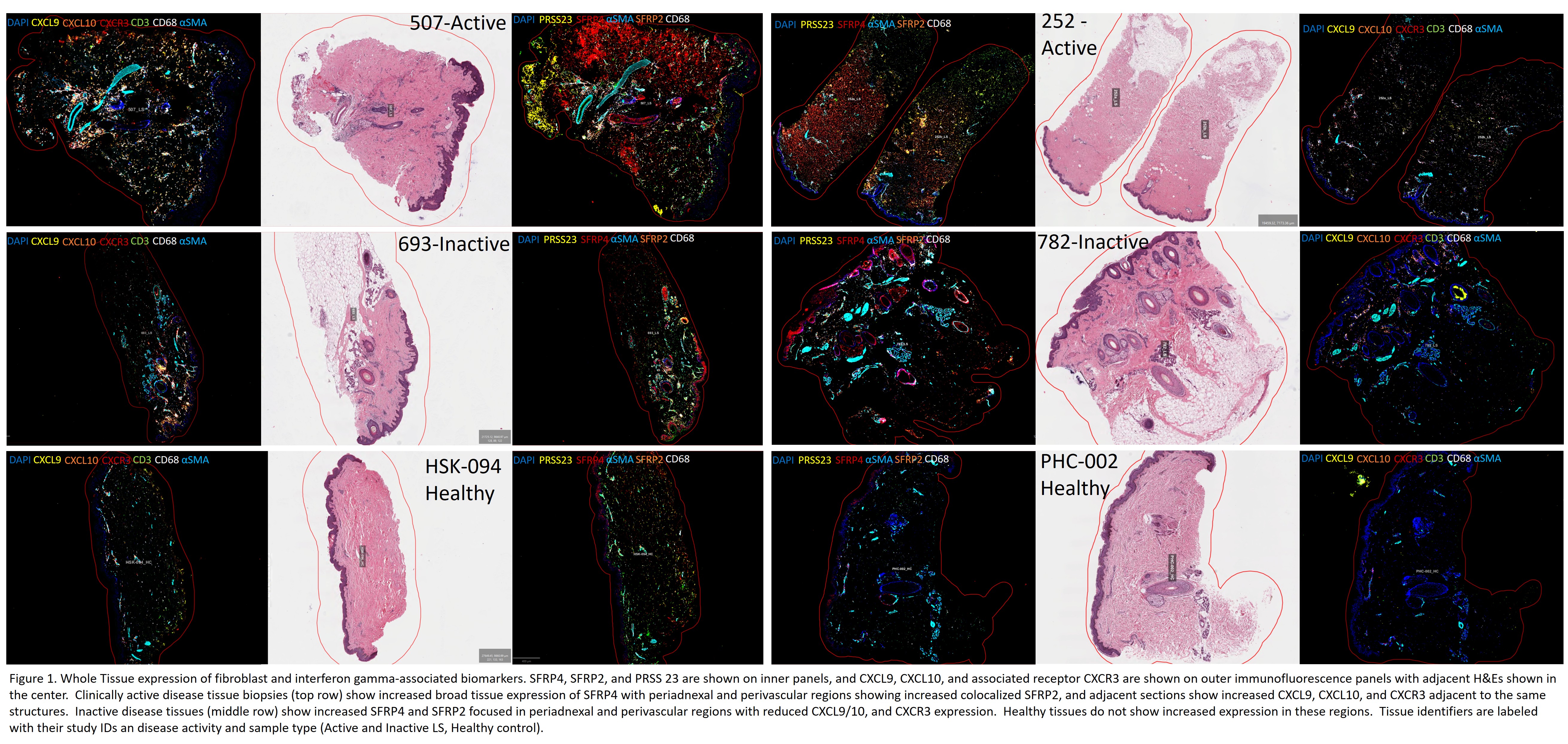
Example Feature of Periadnexal and Perivascular Expression of Secreted Frizzled Proteins and IFNg-related Chemokines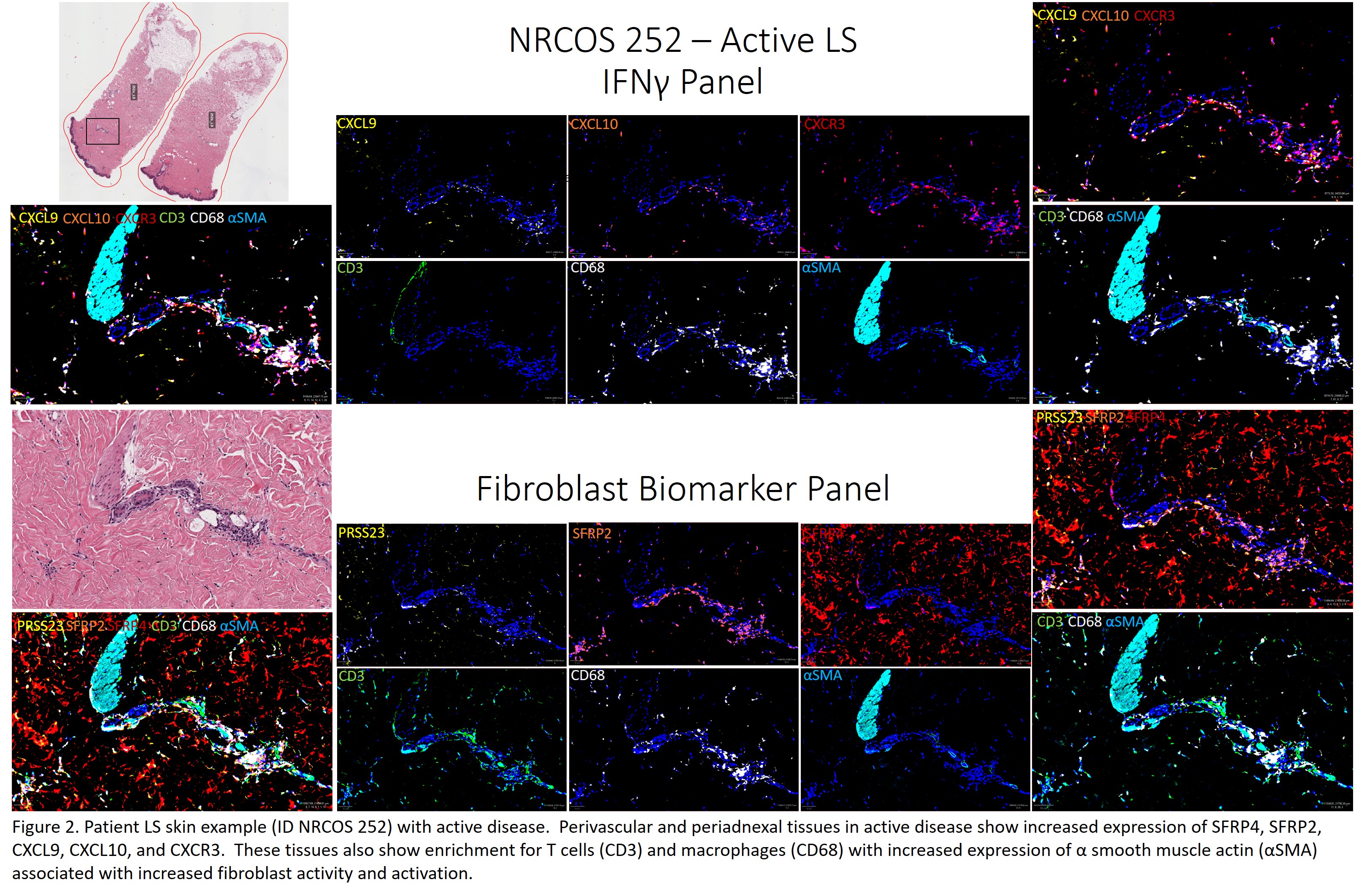
To cite this abstract in AMA style:
Barnett D, Branton S, Esencan D, Lee J, Torok K. Tissue Protein Expression Profiling of Interferon Gamma and Frizzled-Wnt-related Signaling Show Increased Periadnexal and Perivascular Localization in Juvenile Localized Scleroderma Skin [abstract]. Arthritis Rheumatol. 2026; 78 (suppl 3). https://acrabstracts.org/abstract/tissue-protein-expression-profiling-of-interferon-gamma-and-frizzled-wnt-related-signaling-show-increased-periadnexal-and-perivascular-localization-in-juvenile-localized-scleroderma-skin/. Accessed .« Back to 2026 Pediatric Rheumatology Symposium
ACR Meeting Abstracts - https://acrabstracts.org/abstract/tissue-protein-expression-profiling-of-interferon-gamma-and-frizzled-wnt-related-signaling-show-increased-periadnexal-and-perivascular-localization-in-juvenile-localized-scleroderma-skin/