Session Information
Session Type: Poster Session (Monday)
Session Time: 9:00AM-11:00AM
Background/Purpose: Racial disparities exist in healthcare. Research shows that the disproportionate representation of race in educational materials contributes to this disparity. Race is a known risk factor for many diseases, including lupus erythematosus (LE), which predilects for women of color. Given the impact of LE in patients of color, healthcare professionals must learn to recognize the systemic and cutaneous manifestations in this population. We explore whether the skin tones in published images represent the populations that LE affects.
Methods: We conducted a descriptive study, reviewing images from internal medicine, dermatology, and rheumatology textbooks, online image libraries, UpToDate and a comprehensive search of Google Images. We selected textbooks published in the last five years that were available through our university’s online medical library. We identified images by searching for “lupus” and “lupus rash.” We excluded the images if they were in black-and-white, the skin was not clearly visible, or if they were duplicate images. We used the Massey-Martin Skin Color Scale (MMSCS) to grade skin tones from 1 (very light) to 10 (very dark). We sorted the images into four groups: A (MMSCS 1-2), B (MMSCS 3-4), C (MMSCS 5-7), and D (MMSCS 8-10). Each image was scored twice, once by AR, AW or LZ and once by HJ or MM. In cases of discrepancy, HJ or MM broke the tie. Descriptive statistics were used to analyze the data.
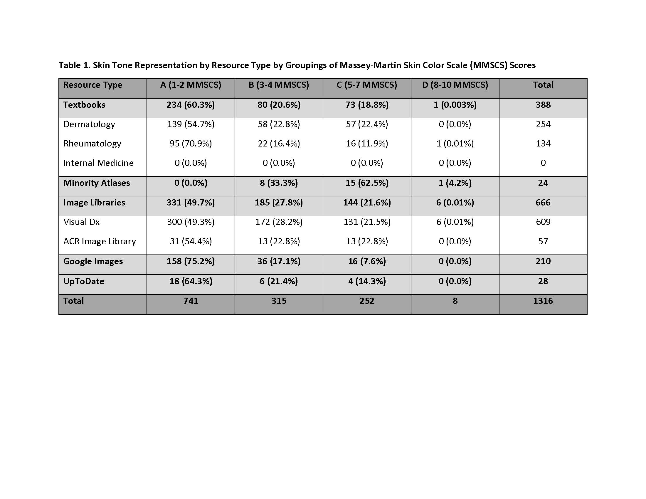
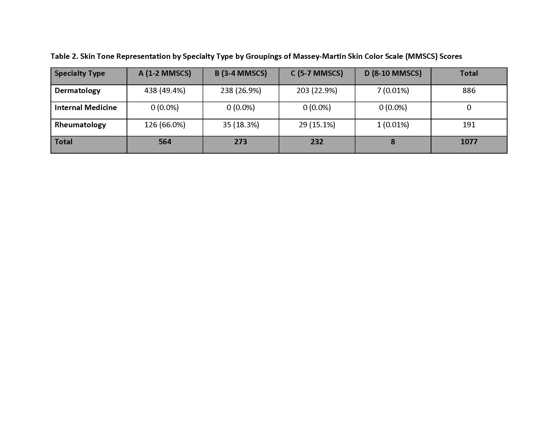
Results: In addition to UpToDate and Google Images, we found 70 dermatology textbooks, 37 rheumatology textbooks, 10 internal medicine textbooks, two minority skin atlases, and two image libraries that met criteria. After exclusions, 1316 images were assessed. The majority (56.3%) of all images represented the skin tones of Group A (Figure 1). By resource type, minority atlases were the most inclusive of skin of color with 66.7% of images categorized in Groups C and D (Table 1). Image libraries provided the next best distribution of skin tones, while Google Images heavily published lighter skin tones (75.2%, Group A). By specialty, dermatology included the greatest variety of skin tones, followed by rheumatology (Table 2). No images in internal medicine texts met inclusion criteria.
Conclusion: We show that lighter skin tones are predominantly represented in a sampling of published images of LE. Ironically, this does not match the clinical demographic of LE. The lack of diversity in medical and online resources has been investigated as a potential causative factor in healthcare disparities. Our study adds to the current data highlighting the paucity of darker skin tones in medical resources. Further research is needed to determine if this affects the confidence and ability of healthcare providers to recognize LE in patients of color.

Lupus Image Study- Figure 1- ACR Abstract

Lupus Image Study- Table 1- ACR Abstract

Lupus Image Study- Table 2- ACR Abstract
To cite this abstract in AMA style:
Rana A, Witt A, Jones H, Mwanthi M, Zickuhr L. The Representation of Skin Tones in Images of Patients with Lupus Erythematosus [abstract]. Arthritis Rheumatol. 2019; 71 (suppl 10). https://acrabstracts.org/abstract/the-representation-of-skin-tones-in-images-of-patients-with-lupus-erythematosus/. Accessed .« Back to 2019 ACR/ARP Annual Meeting
ACR Meeting Abstracts - https://acrabstracts.org/abstract/the-representation-of-skin-tones-in-images-of-patients-with-lupus-erythematosus/
