ACR Meeting Abstracts

ACR Meeting Abstracts

  • Meetings
    • ACR Convergence 2024
    • ACR Convergence 2023
    • 2023 ACR/ARP PRSYM
    • ACR Convergence 2022
    • ACR Convergence 2021
    • ACR Convergence 2020
    • 2020 ACR/ARP PRSYM
    • 2019 ACR/ARP Annual Meeting
    • 2018-2009 Meetings
    • Download Abstracts
  • Keyword Index
  • Advanced Search
  • Your Favorites
    • Favorites
    • Login
    • View and print all favorites
    • Clear all your favorites
  • ACR Meetings

Abstract Number: 1785

The Pathogenic Potential of Choroid Plexus T Cells in Neuropsychiatric Lupus

Erica Moore1, Michelle Huang 1, Cara Reynolds 1 and Chaim Putterman 2, 1Albert Einstein College of Medicine, Bronx, NY, 2Albert Einstein College of Medicine, New York, NY

Meeting: 2019 ACR/ARP Annual Meeting

Keywords: NPSLE and T cells

  • Tweet
  • Email
  • Print
Session Information

Date: Monday, November 11, 2019

Title: 4M094: SLE – Animal Models (1782–1787)

Session Type: ACR Abstract Session

Session Time: 2:30PM-4:00PM

Background/Purpose: The pathogenesis of neuropsychiatric Systemic Lupus Erythematosus (NPSLE) is incompletely understood, but is considered to be complex and multifactorial. Additionally, there is a wide range of possible NPSLE manifestations, including cognitive abnormalities, mood disorders, confusion, psychosis, and seizures. We have recently proposed that the blood-cerebrospinal fluid barrier at the brain choroid plexus (CP), rather than the blood-brain barrier, is the entry site for lymphocytes and other neuroinflammatory mediators into the brain. Indeed, the choroid plexus in the spontaneous MRL/lpr lupus mouse model is heavily infiltrated with predominantly T follicular helper CD4+ T cells and B cells, while  preliminary imaging studies in human NPSLE have similarly revealed inflammation in the region of the CP. However, the function of CP-infiltrating T cells has not been studied as a potential contributor to the pathogenesis of NPSLE.

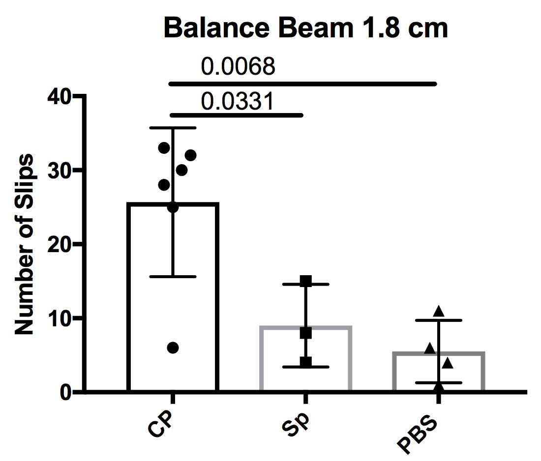
Methods: Six week old female MRL/lpr mice were intracerebroventricularly (ICV) injected with either MRL/lpr choroid plexus CD3+ T cells (n=6), MRL/lpr splenic CD3+ T cells (n=5), or PBS (n=4). The T cells were pooled from the choroid plexus of 16 week old MRL/lpr mice (n=7) and from the spleen of 6 week old MRL/lpr mice (n=2). The cells were stimulated and expanded with anti-CD3/CD28 antibodies (1 ug/mL) and mIL-2 (10 U/mL) for 4 days. About ~200,000 cells were injected into the lateral ventricle from the bregma at AP -0.34 mm, ML -1.0 mm, and depth -2.0 mm. Mice were sacrificed 4 weeks post-injection, with behavioral assessments and histology performed to assess features of NPSLE.

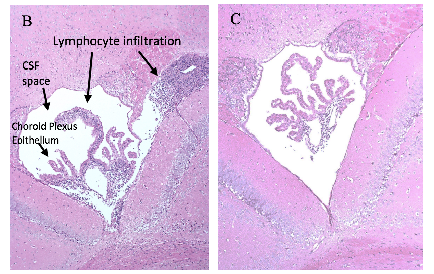
Results: In this pilot study, we found that CP T cell recipient mice had increased cognitive and depressive deficits compared to PBS controls and the MRL/lpr splenic T cell recipient mice. Additionally, the CP T cell recipient mice had a significantly compromised motor function, as assessed by the number of slips while crossing a balance beam (p = 0.0068), when compared to the splenocyte injected groups and the PBS controls (Figure 1A). Histological assessment and immunohistochemical staining revealed an increase in lymphocytic infiltration, the number of proliferating cells (Figure 1B-C), as well as in IgG and complement deposition, in the choroid plexus of CP T recipient mice compared to the PBS group.

Conclusion: Our result indicate that T cells can migrate to and proliferate in the CP following ICV injection, and that brain infiltrating MRL/lpr T cells may be a key contributor to the pathogenesis of NPSLE.

Figure 1A: Motor coordination assessment counting number of slips while crossing a balance beam, CP = choroid plexus, Sp = splenic T cell group, PBS = PBS group

Figure 1B: H&E image of the third ventricle choroid plexus in MRL/lpr CP T cell recipient mouse; Figure 1C: H&E image of the third ventricle choroid plexus in MRL/lpr PBS recipient mouse


Disclosure: E. Moore, None; M. Huang, None; C. Reynolds, None; C. Putterman, Equillium, 5, Equillium, Inc, 2, 5, Exagen, 2.

To cite this abstract in AMA style:

Moore E, Huang M, Reynolds C, Putterman C. The Pathogenic Potential of Choroid Plexus T Cells in Neuropsychiatric Lupus [abstract]. Arthritis Rheumatol. 2019; 71 (suppl 10). https://acrabstracts.org/abstract/the-pathogenic-potential-of-choroid-plexus-t-cells-in-neuropsychiatric-lupus/. Accessed .
  • Tweet
  • Email
  • Print

« Back to 2019 ACR/ARP Annual Meeting

ACR Meeting Abstracts - https://acrabstracts.org/abstract/the-pathogenic-potential-of-choroid-plexus-t-cells-in-neuropsychiatric-lupus/

Advanced Search

Your Favorites

You can save and print a list of your favorite abstracts during your browser session by clicking the “Favorite” button at the bottom of any abstract. View your favorites »

All abstracts accepted to ACR Convergence are under media embargo once the ACR has notified presenters of their abstract’s acceptance. They may be presented at other meetings or published as manuscripts after this time but should not be discussed in non-scholarly venues or outlets. The following embargo policies are strictly enforced by the ACR.

Accepted abstracts are made available to the public online in advance of the meeting and are published in a special online supplement of our scientific journal, Arthritis & Rheumatology. Information contained in those abstracts may not be released until the abstracts appear online. In an exception to the media embargo, academic institutions, private organizations, and companies with products whose value may be influenced by information contained in an abstract may issue a press release to coincide with the availability of an ACR abstract on the ACR website. However, the ACR continues to require that information that goes beyond that contained in the abstract (e.g., discussion of the abstract done as part of editorial news coverage) is under media embargo until 10:00 AM ET on November 14, 2024. Journalists with access to embargoed information cannot release articles or editorial news coverage before this time. Editorial news coverage is considered original articles/videos developed by employed journalists to report facts, commentary, and subject matter expert quotes in a narrative form using a variety of sources (e.g., research, announcements, press releases, events, etc.).

Violation of this policy may result in the abstract being withdrawn from the meeting and other measures deemed appropriate. Authors are responsible for notifying colleagues, institutions, communications firms, and all other stakeholders related to the development or promotion of the abstract about this policy. If you have questions about the ACR abstract embargo policy, please contact ACR abstracts staff at [email protected].

Wiley

  • Online Journal
  • Privacy Policy
  • Permissions Policies
  • Cookie Preferences

© Copyright 2025 American College of Rheumatology