Session Information
Session Type: ACR Poster Session A
Session Time: 9:00AM-11:00AM
Background/Purpose:
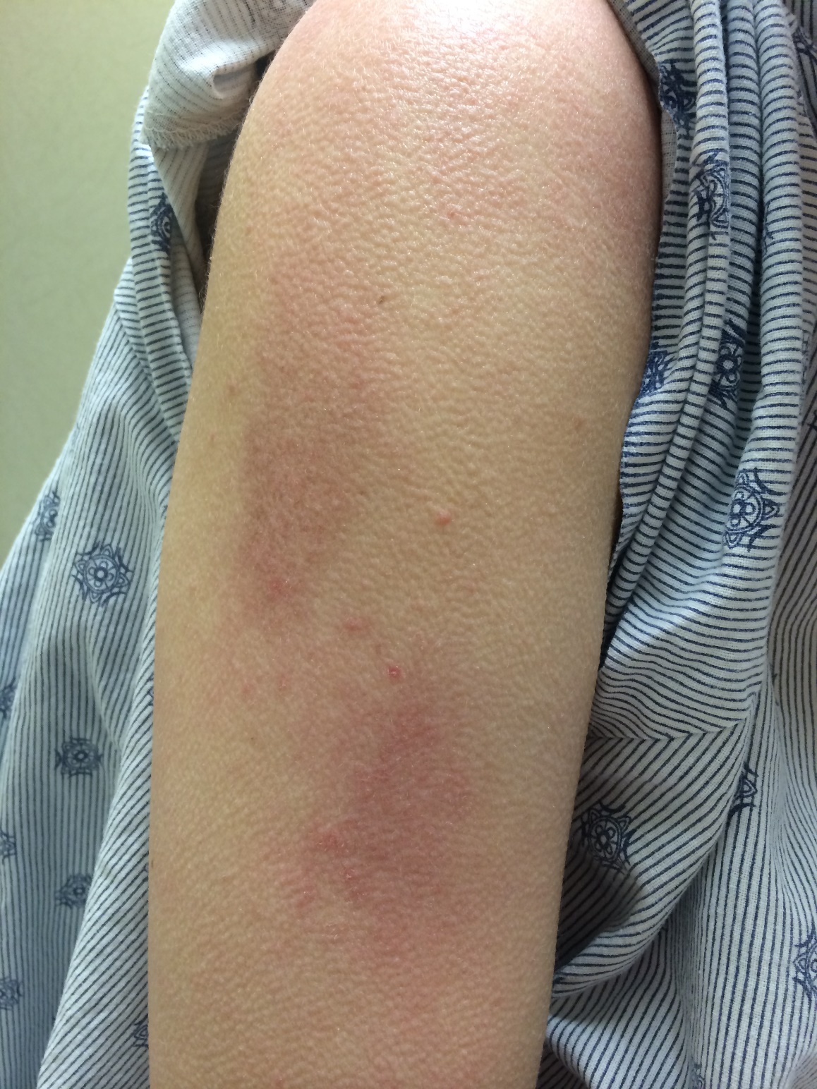
Dermatomyositis (DM) is a subset of idiopathic inflammatory myopathies, characterized by proximal skeletal muscle weakness and skin manifestations. Several skin eruptions have been described in DM patients including Gottron’s papules, Gottron’s sign, heliotrope rash, facial erythema, periungual erythema, shawl sign, V-neck sign, and holster sign. We describe a new skin eruption we have termed the “sleeve sign” observed in 27 patients with DM. To our knowledge, this typical skin finding has not been described before.
Methods:
Two hundred sixty-two patients with a Bohan and Peter diagnosis of definite, probable, or possible DM or a diagnosis of amyopathic DM by Sontheimer’s criteria were evaluated in a systematic fashion including a detailed skin examination with distribution and location of cutaneous findings. All patients underwent clinical evaluation between 2004 and 2015. Demographic and clinical information were retrieved by chart review. Patients’ sera were tested for myositis-specific antibodies using previously validated immunoprecipitation methods.
Results:
Twenty-seven patients (10.3%) had a history of an erythematous to violaceous macule or patch confined to the lateral aspect of the upper arm detected on physical examination. The location was compatible with the contour of the sleeves. Thus, it was labeled the “sleeve sign” (Figure 1).
Twenty-five patients (92.6 %) had dermatomyositis (44.4 % definite, 29.6 % probable, and 18.5 % possible) and 2 patients (7.4%) had amyopathic dermatomyositis. The sera from 22 patients (81.5%) were tested for all DM-specific (anti-TIF-1γ, anti-NXP2, and anti-MDA5) and anti-synthetase autoantibodies (anti-Jo1, anti-PL7/PL12, and anti-EJ/OJ). Table 1 summarizes the demographics, clinical manifestations, and myositis-specific autoantibody profile of the study population.
Conclusion:
A macular erythematous to violaceous rash (sleeve sign) may affect the lateral aspect of the upper arms. Although in our study, the sleeve sign was frequently present with other characteristic DM skin findings, the presence of this rash should raise the suspicion of dermatomyositis in the clinically appropriate setting.
|
Table 1- Demographic, clinical manifestations, and myositis-specific autoantibody profile of patients with sleeve sign |
|
|
Age, mean (SD) |
44.5 (14.3) |
|
Female, number (frequency) |
23 (85.2%) |
|
Race, number (frequency) Caucasian African-American Other |
22 (81.5%) 3 (11.1%) 2 (7.4) |
|
Proximal weakness |
21 (77.8%) |
|
The frequency of the skin findings at the same visit the patient presented with sleeve sign |
|
|
Heliotrope rash |
12 (44.4%) |
|
Malar rash |
14 (51.9%) |
|
Gottron’s sign |
23 (85.2%) |
|
V Sign |
16 (59.3%) |
|
Shawl Sign |
15 (55.6%) |
|
Nail-bed erythema |
17 (63.0%) |
|
Dilated capillary loops |
14 (51.9%) |
|
Holster sign |
7 (25.9%) |
|
Mechanic’s hand |
6 (22.2%) |
|
Calcinosis |
5 (18.5%) |
|
Raynaud’s |
3 (11.1%) |
|
Myositis-specific antibodies |
|
|
Anti-jo1 |
2 (10.5%) |
|
Anti-nxp2 |
4 (22.2%) |
|
Anti-TIF-1γ |
13 (65.0%) |
|
DM-specific or anti-synthetase autoantibody negative |
4 (18.2%) |

To cite this abstract in AMA style:
Lahoutiharahdashti A, Paik JJ, Albayda J, Mammen A, Christopher-Stine L. The “Sleeve Sign”� in Dermatomyositis [abstract]. Arthritis Rheumatol. 2015; 67 (suppl 10). https://acrabstracts.org/abstract/the-aeoesleeve-signae%ef%bf%bd-in-dermatomyositis/. Accessed .« Back to 2015 ACR/ARHP Annual Meeting
ACR Meeting Abstracts - https://acrabstracts.org/abstract/the-aeoesleeve-signae%ef%bf%bd-in-dermatomyositis/