Session Information
Session Type: Abstract Session
Session Time: 3:00PM-4:30PM
Background/Purpose: Ultrasound is a highly valuable imaging modality to study joints in patients with rheumatic diseases. In children, the applicability is hampered because of lack of normative data, and especially data adjusted to the age of the growing child. The purpose of this study is to examine structural sonographic features of joints and tendons in healthy children and adolescents amongst several age groups, and develop a set of normative data, in order to establish reference values.
Methods: 500 healthy volunteers (age 0 – 18 years) were scanned according to a predefined scanning protocol including the right shoulder, hip, knee, ankle, first metatarsophalangeal joint (MTP1), elbow, wrist and second metacarpophalangeal joint (MCP2). The following structures and variables were captured: synovial capsular distention at the acetabulofemoral, the supra- and parapatellar, the tibiotalar, the MTP1 joint, the lateral and anterior radiohumeral, the posterior fossa of the elbow, the radio-lunate, lunate-capitatum and capitatum-metacarpal and the MCP2 joint recesses; the diameter of the extensor digitorum communis, extensor carpi ulnaris, the patellar and the biceps tendon; and cartilage thickness at the femoral head, the femoral trochlea, the talar dome, head of MCP2 and MTP1. Unpaired T-test, paired T-test, correlation analyses and regression analysis were performed on the collected data. The significance level (α) was set at 0.1% (p < 0.001) in all analyses. Growth charts are used to build reference values of the outcomes of interest with respect to age. Nonparametric quantile regression with a penalty on the coefficients and an estimated smoothing parameter was applied using the R-packages quantreg and quantregGrowth. Interobserver reliability exercise and analysis was performed.
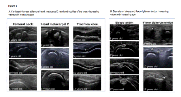
Results: Images of 195 male and 305 female volunteers were collected. Interobserver reliability was excellent for new and experienced sonographers (all intraclass coefficients of correlation > 0.8). In all joints, cartilage diminished markedly as children age (figure 1) and cartilage of boys was significantly thicker compared to girls in most of the joints. In addition, cartilage also became thinner as children’s height and weight increased. Capsular distention ( > 0mm) was uncommon in the ankle, wrist and MCP2 (resp. in 3, 6, and 3% of cases). It was more common in the suprapatellar and parapatellar knee, MTP1 and posterior recess of the elbow (resp. in 34, 32, 46, and 39% of cases). In the hip, capsular distention was always present. Age is only moderately associated to capsular distention at the hip, knee and MTP1 joint, and only poorly at the ankle, elbow, and wrist. Upon ageing, the diameter of tendons thickened (figure 1). Growth curves and tables are available for each variable (figure 2 shows growth charts of acetabulofemoral recess and thickness of cartilage at MTP1).
Conclusion: Reference values for sonographic values of cartilage thickness, capsular distention and diameters of tendons at several joints in several age groups were established from 500 healthy children, aged between 0 and 18 years. The results of this study are indispensable for the interpretation of ultrasonographic findings in children with inflammatory arthritis.
To cite this abstract in AMA style:
Wittoek R, Decock C, Dewaele N, Arnold L, baeyens P, Deschrijver I, Pardaens L, Raftakis I, Renson T, Thooft A, Rinkin C, Vanhaverbeke T, Verbist C. Structural Ultrasound Features of Joints and Tendons of Healthy Children: Development of Normative Data [abstract]. Arthritis Rheumatol. 2022; 74 (suppl 9). https://acrabstracts.org/abstract/structural-ultrasound-features-of-joints-and-tendons-of-healthy-children-development-of-normative-data/. Accessed .« Back to ACR Convergence 2022
ACR Meeting Abstracts - https://acrabstracts.org/abstract/structural-ultrasound-features-of-joints-and-tendons-of-healthy-children-development-of-normative-data/


