ACR Meeting Abstracts

ACR Meeting Abstracts

  • Meetings
    • ACR Convergence 2024
    • ACR Convergence 2023
    • 2023 ACR/ARP PRSYM
    • ACR Convergence 2022
    • ACR Convergence 2021
    • ACR Convergence 2020
    • 2020 ACR/ARP PRSYM
    • 2019 ACR/ARP Annual Meeting
    • 2018-2009 Meetings
    • Download Abstracts
  • Keyword Index
  • Advanced Search
  • Your Favorites
    • Favorites
    • Login
    • View and print all favorites
    • Clear all your favorites
  • ACR Meetings

Abstract Number: 576

Spinal Bone Formation as Assessed by Low-Dose CT Scan in Patients with Radiographic Axial Spondyloarthritis – Comparison of the Progression Observed in Vertebrae and Facet Joints

Rosalinde Stal1, Floris van Gaalen 1, Alexandre Sepriano 1, Jürgen Braun 2, Monique Reijnierse 1, Désirée van der Heijde 1 and Xenofon Baraliakos 3, 1Leiden University Medical Center, Leiden, Netherlands, 2Rheumazentrum Ruhrgebiet/Ruhr University, Herne, Germany, 3Rheumazentrum Ruhrgebiet-Ruhr-University Bochum, Herne, Germany, Herne, Germany

Meeting: 2019 ACR/ARP Annual Meeting

Keywords: axial spondyloarthritis and Imaging

  • Tweet
  • Click to email a link to a friend (Opens in new window) Email
  • Click to print (Opens in new window) Print
Session Information

Date: Sunday, November 10, 2019

Title: Spondyloarthritis Including Psoriatic Arthritis – Clinical Poster I: Axial Spondyloarthritis, Clinical Features

Session Type: Poster Session (Sunday)

Session Time: 9:00AM-11:00AM

Background/Purpose: By using low dose computed tomography (ldCT) in patients with radiographic axial spondyloarthritis (r-axSpA) we have recently shown that there is substantial progression of new bone formation in the facet joints within two years. We now aimed to assess the added value of scoring progression of whole spine facet joint ankylosis in addition to  whole spine syndesmophytes in the quantification of spinal bone formation by ldCT.

Methods: In an observational cohort, patients with r-axSpA (≥1 inflammatory lesion on spinal MRI and 1-18 syndesmophytes in cervical and lumbar spine on conventional radiography) underwent whole spine ldCT (circa 4 mSv) at baseline and after 2 years. Two trained readers independently assessed paired ldCTs, blinded to chronology. Left and right facet joints from C2-S1, with the exception of C5-T2 due to low visibility, were scored as ankylosis present (1) or absent (0). Syndesmophytes were scored on a scale 0-552 according the Computed Tomography Syndesmophyte Score (CTSS)[1]. Average scores of the readers were used. Patients were included if at baseline they had < 80% ankylosed facet joints and < 80% bridged syndesmophytes on ldCT and had scores from both readers for both timepoints. Change scores are calculated as the total change in the whole spine per patient. The proportion of patients with a change score >0.5 and >SDC are presented and in addition net progression, defined as number of patients with change >0.5 minus number of patients with change < -0.5 divided by the total number of patients. Percentage agreement between progression in facet ankylosis and syndesmophyte formation is presented and a cumulative probability plot is shown.

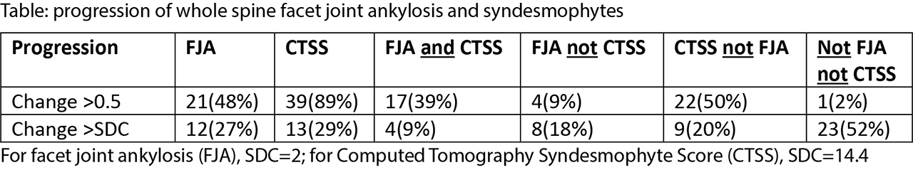
Results: A total of 44 patients was included in the analysis (mean age 49.4, 84% male, 79% HLA-B27+). At the group level, net progression of facet joint ankylosis was 43% (19/44) and net progression of syndesmophytes was 82% (36/44). The proportion of patients with progression is given in the Table. The percentage of patients with progression of facet joint ankylosis >0.5 was 48% and with syndesmophyte progression >0.5 was 89%. Using a more conservative cut-off for progression ( >SDC), the percentage of patients with progression of facet joint ankylosis was 27% and syndesmophyte progression was seen in 29%. Bone formation in the facet joints only occurred in 9% and 18% of the patients, and syndesmophytes progression only  in 50% and 20% of the patients for the cut-offs of 0.5 and SDC, respectively. The figure shows that there is a tendency that patients with more facet joint ankylosis progression also had more syndesmophyte progression. However, in a number of patients these processes occurred independently.

Conclusion: These data show that scoring facet joint ankylosis in addition to syndesmophyte formation is useful to get full insight in the progression of spinal bone formation in patients with r-axSpA.

[1] de Bruin, F., et al. (2018). “Development of the CT Syndesmophyte Score (CTSS) in patients with ankylosing spondylitis: data from the SIAS cohort.” Annals of the rheumatic diseases 77(3): 371-377


Disclosure: R. Stal, None; F. van Gaalen, None; A. Sepriano, None; J. Braun, Abbott, 2, 5, AbbVie, 2, 5, 6, 8, Amgen, 2, 5, 8, Baxter, 2, 5, 8, Biogen, 2, 8, 9, BMS, 2, 5, 8, Boehringer, 2, 5, 8, Bristol-Myers Squibb, 2, 5, Celgene, 2, 3, 5, 8, Celltrion, 2, 5, 8, Centocor, 2, 5, 8, Chugai, 2, 5, 8, Eli Lilly and Company, 2, 5, 8, Hexal, 2, 5, 8, Janssen, 2, 5, 8, Johnson & Johnson, 2, 5, Medac, 2, 5, 8, MSD, 2, 5, MSD (Schering-Plough), 2, 5, 8, Mundipharma, 2, 5, 8, Mylan, 2, 5, 8, Novartis, 2, 5, 8, Pfizer, 2, 5, Pfizer (Wyeth, Hospira), 2, 5, 8, Roche, 2, 5, 8, Sanofi-Aventis, 2, 5, 8, UCB Pharma, 2, 5, 8; M. Reijnierse, None; D. van der Heijde, AbbVie, 5, AbbVie, Amgen, Astellas, AstraZeneca, BMS, 5, Amgen, 5, Astellas, 5, 9, Astellas Pharma, 5, AstraZeneca, 5, BMS, 5, Boehringer Ingelheim, 5, Boehringer Ingelheim, Celgene, Daiichi, Eli-Lilly, Galapagos, Gilead, GSK, Janssen, Merck, Novartis, Pfizer, Regeneron, Roche, Sanofi, Takeda, UCB, 5, Boehringer-Ingelheim, 5, Bristol-Myers Squibb, 5, Celgene, 5, Daiichi, 5, 9, Daiichi Sankyo, 5, Director of Imaging Rheumatology, 6, Director of Imaging Rheumatology bv, 9, Eli Lilly, 5, Eli Lilly and Company, 5, Eli-Lilly, 5, Galapagos, 5, Gilead, 5, Gilead Sciences, Inc., 5, GlaxoSmithKline, 5, Glaxo-Smith-Kline, 5, GSK, 5, 8, Imaging Rheumatology bv, 9, Imaging Rheumatology BV, 9, Imaging Rheumatology bv., 9, Janssen, 5, 8, Janssen Pharmaceutica, 5, Merck, 5, 8, Novartis, 5, 8, Pfizer, 5, 8, Pfizer Inc, 5, Regeneron, 5, 8, Rheumatology bv, 4, 9, Roche, 5, 8, Sanofi, 5, 8, Takeda, 5, 8, Takeda Pharmaceutical Company, 5, UCB, 5, 8, UCB Pharma, 5; X. Baraliakos, AbbVie, 2, 5, 8, Abbvie, 2, 5, 8, BMS, 2, 5, 8, 9, Bristol-Myers Squibb, 2, 5, 8, Celgene, 2, 5, 8, 9, Chugai, 2, 5, 8, 9, Janssen, 2, 5, 8, 9, Lilly, 2, 8, 9, Merck, 2, 5, 8, MSD, 2, 5, 8, 9, Novartis, 2, 5, 8, 9, Novatis, 2, 5, 8, Pfizer, 2, 5, 8, 9, UCB, 2, 5, 8, 9, UCB Pharma, 2, 5, 8, Werfen, 2, 5, 8.

To cite this abstract in AMA style:

Stal R, van Gaalen F, Sepriano A, Braun J, Reijnierse M, van der Heijde D, Baraliakos X. Spinal Bone Formation as Assessed by Low-Dose CT Scan in Patients with Radiographic Axial Spondyloarthritis – Comparison of the Progression Observed in Vertebrae and Facet Joints [abstract]. Arthritis Rheumatol. 2019; 71 (suppl 10). https://acrabstracts.org/abstract/spinal-bone-formation-as-assessed-by-low-dose-ct-scan-in-patients-with-radiographic-axial-spondyloarthritis-comparison-of-the-progression-observed-in-vertebrae-and-facet-joints/. Accessed .
  • Tweet
  • Click to email a link to a friend (Opens in new window) Email
  • Click to print (Opens in new window) Print

« Back to 2019 ACR/ARP Annual Meeting

ACR Meeting Abstracts - https://acrabstracts.org/abstract/spinal-bone-formation-as-assessed-by-low-dose-ct-scan-in-patients-with-radiographic-axial-spondyloarthritis-comparison-of-the-progression-observed-in-vertebrae-and-facet-joints/

Advanced Search

Your Favorites

You can save and print a list of your favorite abstracts during your browser session by clicking the “Favorite” button at the bottom of any abstract. View your favorites »

All abstracts accepted to ACR Convergence are under media embargo once the ACR has notified presenters of their abstract’s acceptance. They may be presented at other meetings or published as manuscripts after this time but should not be discussed in non-scholarly venues or outlets. The following embargo policies are strictly enforced by the ACR.

Accepted abstracts are made available to the public online in advance of the meeting and are published in a special online supplement of our scientific journal, Arthritis & Rheumatology. Information contained in those abstracts may not be released until the abstracts appear online. In an exception to the media embargo, academic institutions, private organizations, and companies with products whose value may be influenced by information contained in an abstract may issue a press release to coincide with the availability of an ACR abstract on the ACR website. However, the ACR continues to require that information that goes beyond that contained in the abstract (e.g., discussion of the abstract done as part of editorial news coverage) is under media embargo until 10:00 AM ET on November 14, 2024. Journalists with access to embargoed information cannot release articles or editorial news coverage before this time. Editorial news coverage is considered original articles/videos developed by employed journalists to report facts, commentary, and subject matter expert quotes in a narrative form using a variety of sources (e.g., research, announcements, press releases, events, etc.).

Violation of this policy may result in the abstract being withdrawn from the meeting and other measures deemed appropriate. Authors are responsible for notifying colleagues, institutions, communications firms, and all other stakeholders related to the development or promotion of the abstract about this policy. If you have questions about the ACR abstract embargo policy, please contact ACR abstracts staff at [email protected].

Wiley

  • Online Journal
  • Privacy Policy
  • Permissions Policies
  • Cookie Preferences

© Copyright 2025 American College of Rheumatology