Session Information
Date: Sunday, November 17, 2024
Title: Vasculitis – Non-ANCA-Associated & Related Disorders Poster II
Session Type: Poster Session B
Session Time: 10:30AM-12:30PM
Background/Purpose: Assessing disease activity in Takayasu arteritis (TA) is a significant challenge, and there is no consensus on evaluating inflammatory activity (IA). Previous studies have suggested that interleukin 6 (IL-6) is a central cytokine in the pathophysiology of TA and is the biomarker that best reflects IA in the arterial wall. Here, we compare serum interleukin IL-6 with activity by Indian Takayasu Clinical Arteritis Score (ITAS.A) and CT angiography (CTA) scans.
Methods: In this transversal study, we examined 43 TA patients between January 2022 and July 2023. All patients met the 2022 ACR Criteria for TA. We meticulously assessed clinical, laboratory, and imaging data. IL-6 was measured using the ELISA Invitrogen kit, and C-reactive protein (CRP) was evaluated in the same blood sample. Complete CTA scans were considered if performed within a maximum period of one year from the blood collection. We defined IA if ITAS.A-CRP > 5 (ITAS-IA) and if wall thickening with post-contrast enhancement was reported in at least one selected vessel in CTA (CTA-IA). We then performed linear regression with continuous IL-6 levels and categorical variables ITAS-IA and CTA-IA to compare the results.
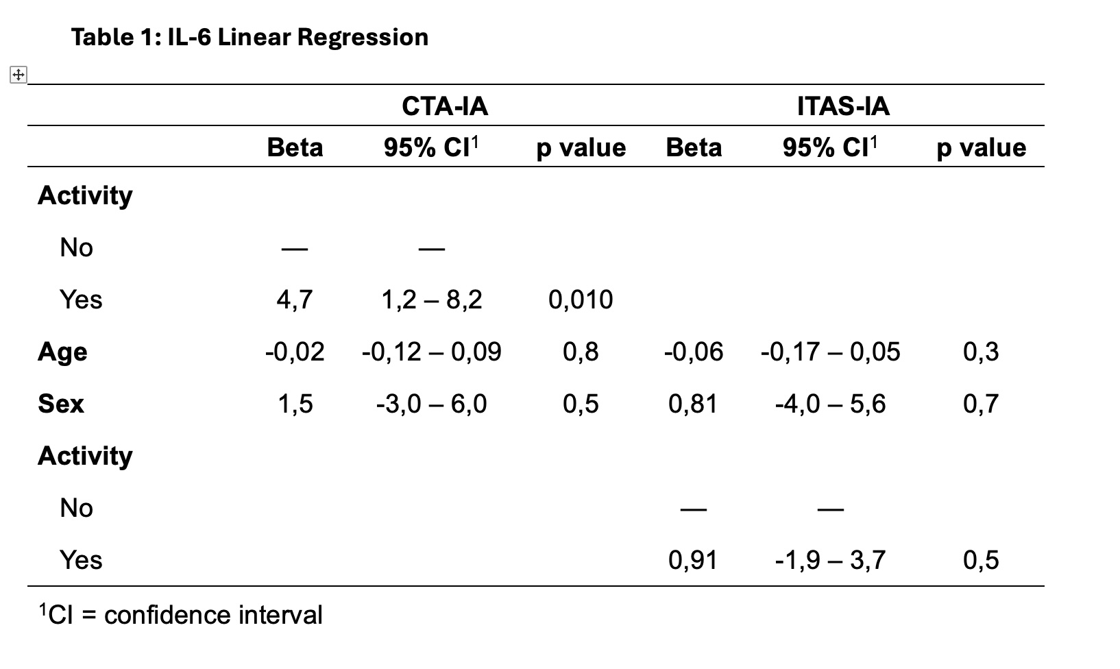
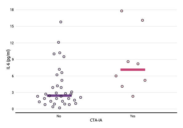
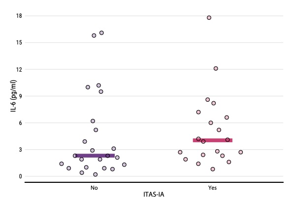
Results: Median age at TA diagnosis was 33 years, 39 were females (90.7%), and Numano V Classification occurred in 28 patients (65.1%). The median CRP was 12.3mg/L (6.8-27.4), and the median IL-6 was 5,2 pg/ml (2,75-9,05). Prednisone was used in 41,8% of cases with a median dose of 10mg (5-20mg), MTX in 22 (51,2%), two patients used anti-TNF (4,6%), and none used tocilizumab. ITAS-IA occurred in 20 patients (46,5%) and CTA-IA in 8 (18,6%). CTA-IA was associated with the IL-6 values of 4.7 greater than non-active individuals by image (95% Confidence interval 1.2 – 8.2 with p: 0,010), a pattern not seen when ITAS-A assessed activity.
Conclusion: Serum IL-6 was higher in patients with active disease by CTA. The combination of IL-6 and image parameters is a potential tool to define the inflammatory status. Careful correlation with clinical and other laboratory and radiological assessments is needed. However, further research is needed to develop composite disease activity scores that integrate serum biomarkers with imaging, thereby advancing our understanding and management of this complex condition.
To cite this abstract in AMA style:
Souza Pedreira A, Nunez Costa G, da Silva Cendon Duran C, Paz A, Matos A, Silva De Oliveira I, Barreto Santiago M. Serum IL-6 Levels Correlate with Inflammatory Activity in Computerized Tomography Angiography Scans in Takayasu Arteritis [abstract]. Arthritis Rheumatol. 2024; 76 (suppl 9). https://acrabstracts.org/abstract/serum-il-6-levels-correlate-with-inflammatory-activity-in-computerized-tomography-angiography-scans-in-takayasu-arteritis/. Accessed .« Back to ACR Convergence 2024
ACR Meeting Abstracts - https://acrabstracts.org/abstract/serum-il-6-levels-correlate-with-inflammatory-activity-in-computerized-tomography-angiography-scans-in-takayasu-arteritis/


