Session Information
Session Type: ACR Abstract Session
Session Time: 2:30PM-4:00PM
Background/Purpose: Synovial neoangiogenesis is an early and crucial event to promote the development of the hyperplasic proliferative pathologic synovium in rheumatoid arthritis (RA). A recent microarray analysis revealed a differential expression of semaphorin family in RA endothelial cells (ECs), which is known to contribute to angiogenesis and inflammation. Our aim was to study the potential implication of semaphorins in RA pathogenesis.
Methods: mRNA levels of class 3 and 4 Semaphorins (SEMA) SEMA3A, SEMA3E, SEMA4A and SEMA4E, as well as their receptors PlexinD1 (PLXND1) and Neuropillin-1 (NRP1), were measured by real-time quantitative PCR in RA (n=15) and control (n=15) ECs. Protein expression of these 4 semaphorins and their receptors was evaluated in ECs of 10 RA patients and controls by western blot, and in the synovial tissue of 10 RA patients and 5 controls by immunohistochemistry and immunofluorescence. Serum concentrations of these 4 semaphorins were measured by sandwich ELISA in a cohort of 130 patients with RA (85% women, mean age: 58 ± 12 years and mean disease duration: 11 ± 12 years) and 30 age- (56 ± 10 years) and sex- (82% women) matched controls.
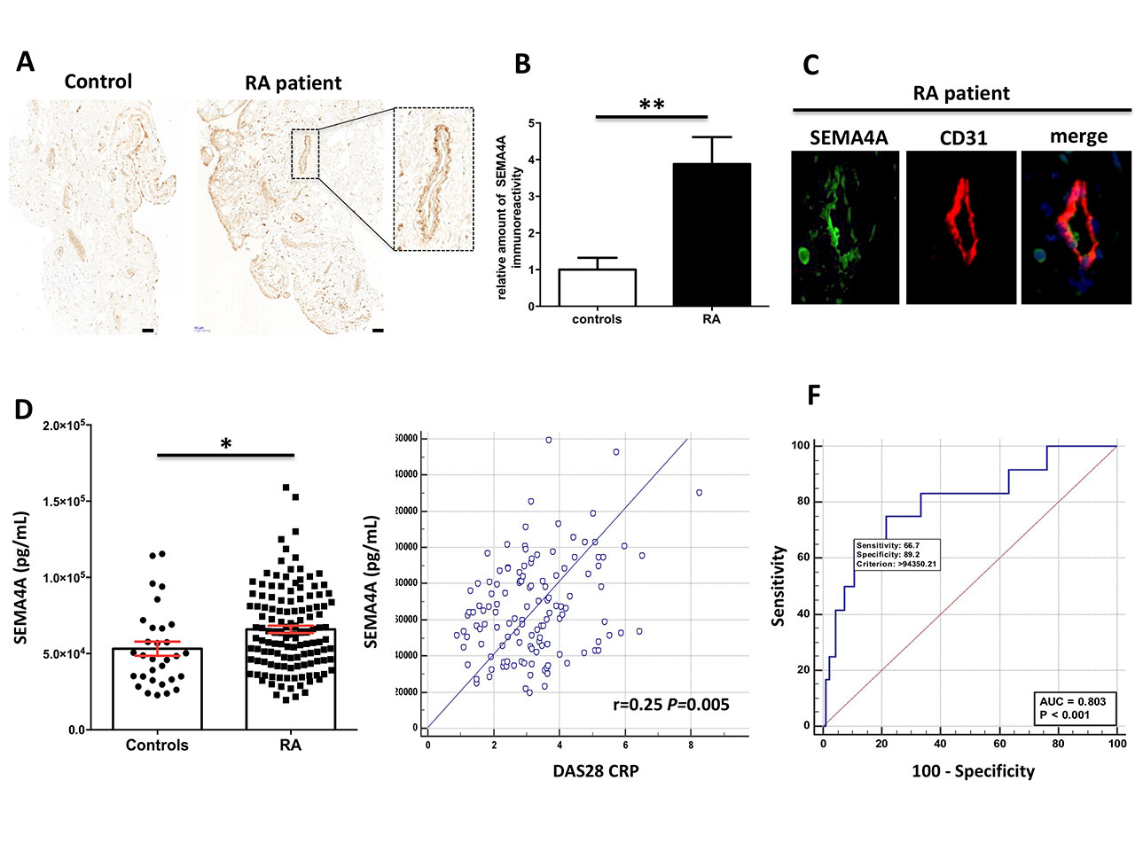
Results: Results:SEMA4A, PLXND1 and NRP1 mRNA and protein levels were markedly increased in RA ECs. The expression of SEMA4A (Figure 1A-B), SEMA3A, SEMA3E, PLXND1 and NRP1 was strikingly increased in the synovial tissue of RA patients. Confocal microscopy with double labeling for CD31 confirmed the prominent endothelial expression of these class 3 and 4 semaphorins and their receptors (Figure 1C). Serum levels of SEMA4A (Figure 1D) and SEMA3E were significantly increased in patients with RA compared to controls. Conversely, SEMA3A serum levels were strikingly lower in patients with RA compared to controls. SEMA4A (Figure 1E), SEMA3A, SEMA3E and SEMA4D serum levels correlated with markers of joint and systemic inflammation (swollen joint count, ESR, CRP, synovial vascularization assessed by power Doppler), validated composite score measuring disease activity (DAS28 and DAS28-CRP) as well as circulating proangiogenic markers (VEGF, Tie2, VCAM-1, IL-8). The diagnostic value of SEMA4A to identify patients with an “inflammatory profile” (DAS28 >3.2, CRP >10 mg/L and increased power Doppler global arthritis score) was defined by an area under curve (AUC) of 0.80 (P< 0.001) (Figure 1F). In the 52/130 patients (40%) in remission or with low disease activity defined by a DAS28< 3.2, defined by the persistence of synovial hyperemia detected on at least one joint, SEMA4A and SEMA4D detected residual infraclinical disease activity with an AUC of 0.70 (P=0.008 and P=0.012, respectively).
Conclusion: Gene expression profiling of ECs identified semaphorins as potential biomarkers and therapeutic candidates in RA. Class 3 and 4 semaphorins are overexpressed in ECs, synovial vessels and the serum of patients with RA. They also correlated with validated markers of inflammation and angiogenesis. Thus, semaphorins might be novel and appealing EC-derived inflammatory and proangiogenic targets in RA.
To cite this abstract in AMA style:
Avouac J, Pezet S, Vandebeuque E, Allanore Y. Semaphorins: From Angiogenesis to Inflammation in Rheumatoid Arthritis [abstract]. Arthritis Rheumatol. 2019; 71 (suppl 10). https://acrabstracts.org/abstract/semaphorins-from-angiogenesis-to-inflammation-in-rheumatoid-arthritis/. Accessed .« Back to 2019 ACR/ARP Annual Meeting
ACR Meeting Abstracts - https://acrabstracts.org/abstract/semaphorins-from-angiogenesis-to-inflammation-in-rheumatoid-arthritis/

