ACR Meeting Abstracts

ACR Meeting Abstracts

  • Meetings
    • ACR Convergence 2024
    • ACR Convergence 2023
    • 2023 ACR/ARP PRSYM
    • ACR Convergence 2022
    • ACR Convergence 2021
    • ACR Convergence 2020
    • 2020 ACR/ARP PRSYM
    • 2019 ACR/ARP Annual Meeting
    • 2018-2009 Meetings
    • Download Abstracts
  • Keyword Index
  • Advanced Search
  • Your Favorites
    • Favorites
    • Login
    • View and print all favorites
    • Clear all your favorites
  • ACR Meetings

Abstract Number: 1008

Segmentation and Quantification of Bone Erosions in the Hands of Patients with Rheumatoid Arthritis Using High Resolution Computed Tomography

Dominique Toepfer1, Stephanie Finzel2, Oleg Museyko1, Klaus Engelke3 and Georg A. Schett2, 1Institute of Medical Physics, University of Erlangen-Nuremberg, Erlangen, Germany, 2Dept of Medicine 3, Rheumatology and Clinical Immunology, University of Erlangen-Nuremberg, Erlangen, Germany, 3Institute of Medical Physics, University of Erlangen, University of Erlangen-Nuremberg, Erlangen, Germany

Meeting: 2012 ACR/ARHP Annual Meeting

Keywords: imaging techniques and rheumatoid arthritis (RA)

  • Tweet
  • Click to email a link to a friend (Opens in new window) Email
  • Click to print (Opens in new window) Print
Session Information

Title: Imaging of Rheumatic Diseases: Magnetic Resonance Imaging, Computed Tomography and X-ray

Session Type: Abstract Submissions (ACR)

Background/Purpose: Bone erosions are important for diagnosis and monitoring of disease activity in RA. However, semi-quantitative scoring schemes may be inadequate for a true 3D quantification of size and shape. Recently high-resolution peripheral quantitative CT (HR-pQCT) with an isotropic spatial resolution of about 120µm has been used for a semiquantitative assessment of erosion volume in the metacarpophalangeal (MCP) joints. Here we developed a highly automated 3D analysis technique to more accurately quantify volume, shape and surface of erosions and to increase precision. 

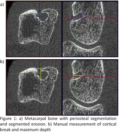
Methods: In the MCP joints of the second to forth digit of  18 patients 80 slices distally and 242 slices proximally to the surface of the third metacarpal head were scanned (XtremeCT, Scanco Switzerland; isotropic voxel size 82µm). Erosions were quantified as follows: After segmenting the periosteal surface the user identified each erosion by manually placing a seed point. The erosions were then automatically segmented by a 3D level-set algorithm (Fig. 1a) with the option of operator corrections. Erosion volume (Vol), surface area (SA), and sphericity (SP), a parameter describing the shape deviation from a perfect sphere, were determined. In addition manual measurements were carried out. Here the erosion volume was approximated by a half-ellipsoid constructed from the surface area of the cortical break and the maximum erosion depth perpendicular to it (Fig. 1b). In order to compare both methods the lengths measurements obtained from the manual technique were also determined during the automated 3D analysis.

Results: 32 erosions were assessed in the 18 datasets with a mean/min/max Vol of 9.66mm³/0.37mm³/54.7mm³ (automated 3D analysis). Inter-operator precision errors (3 operators, root mean squares coefficients of variation (RMSCV)/RMS standard deviation (RMSSD)) were 7.8%/0.8 mm³, 10.4%/3.8 mm2 and 4.9%/0.02 for Vol, SA and SP, respectively. Excluding 18 erosions, in which operator interactions were performed, decreased the errors by about a factor of 3. Correlation between the manual analysis and the length measurements obtained from the automated 3D analysis was r = 0.87, however, correlation with the Vol obtained from the full 3D analysis was r = 0.39, indicating that a simplistic approximation of erosion volume may not capture the full shape information.

Conclusion: We developed a new precise full 3D characterization of bone erosions that may help improving the assessment of disease activity and treatment efficacy. Precision errors depend on the degree of user interaction that may be necessary to correct the automated segmentation, which is more frequent in erosions with large cortical breaks. Manual measurements are less impacted by image quality, such as motion artifacts; however, the approximation of erosion volume by a half-ellipsoid underestimates the true erosion volume. The clinical evaluation of this method is currently being performed.


Disclosure:

D. Toepfer,
None;

S. Finzel,
None;

O. Museyko,
None;

K. Engelke,
None;

G. A. Schett,
None.

  • Tweet
  • Click to email a link to a friend (Opens in new window) Email
  • Click to print (Opens in new window) Print

« Back to 2012 ACR/ARHP Annual Meeting

ACR Meeting Abstracts - https://acrabstracts.org/abstract/segmentation-and-quantification-of-bone-erosions-in-the-hands-of-patients-with-rheumatoid-arthritis-using-high-resolution-computed-tomography/

Advanced Search

Your Favorites

You can save and print a list of your favorite abstracts during your browser session by clicking the “Favorite” button at the bottom of any abstract. View your favorites »

All abstracts accepted to ACR Convergence are under media embargo once the ACR has notified presenters of their abstract’s acceptance. They may be presented at other meetings or published as manuscripts after this time but should not be discussed in non-scholarly venues or outlets. The following embargo policies are strictly enforced by the ACR.

Accepted abstracts are made available to the public online in advance of the meeting and are published in a special online supplement of our scientific journal, Arthritis & Rheumatology. Information contained in those abstracts may not be released until the abstracts appear online. In an exception to the media embargo, academic institutions, private organizations, and companies with products whose value may be influenced by information contained in an abstract may issue a press release to coincide with the availability of an ACR abstract on the ACR website. However, the ACR continues to require that information that goes beyond that contained in the abstract (e.g., discussion of the abstract done as part of editorial news coverage) is under media embargo until 10:00 AM ET on November 14, 2024. Journalists with access to embargoed information cannot release articles or editorial news coverage before this time. Editorial news coverage is considered original articles/videos developed by employed journalists to report facts, commentary, and subject matter expert quotes in a narrative form using a variety of sources (e.g., research, announcements, press releases, events, etc.).

Violation of this policy may result in the abstract being withdrawn from the meeting and other measures deemed appropriate. Authors are responsible for notifying colleagues, institutions, communications firms, and all other stakeholders related to the development or promotion of the abstract about this policy. If you have questions about the ACR abstract embargo policy, please contact ACR abstracts staff at [email protected].

Wiley

  • Online Journal
  • Privacy Policy
  • Permissions Policies
  • Cookie Preferences

© Copyright 2025 American College of Rheumatology