ACR Meeting Abstracts

ACR Meeting Abstracts

  • Meetings
    • ACR Convergence 2024
    • ACR Convergence 2023
    • 2023 ACR/ARP PRSYM
    • ACR Convergence 2022
    • ACR Convergence 2021
    • ACR Convergence 2020
    • 2020 ACR/ARP PRSYM
    • 2019 ACR/ARP Annual Meeting
    • 2018-2009 Meetings
    • Download Abstracts
  • Keyword Index
  • Advanced Search
  • Your Favorites
    • Favorites
    • Login
    • View and print all favorites
    • Clear all your favorites
  • ACR Meetings

Abstract Number: 3084

Progression of Lupus Pathology Is Correlative with Cardiac Magnetic Resonance Imaging Abnormalities, Diminished Function, and Inflammatory Histopathology in an Animal Model

Nicholas Young1, Jeffrey Hampton1, Saba Aqel1, Jessica Pyles2, Anna Bratasz2, Anuradha Kalyanasundaram3, Wael N. Jarjour4 and Stacy P. Ardoin5, 1Immunology and Rheumatology, The Ohio State University Medical Center, Columbus, OH, 2Small Animal Imaging Core, The Ohio State University Medical Center, Columbus, OH, 3Physiology and Cell Biology, The Ohio State University Medical Center, Columbus, OH, 4Department of Internal Medicine, Division of Immunology and Rheumatology, The Ohio State University Medical Center, Columbus, OH, 5Pediatric & Adult Rheumatology, Ohio State University, Columbus, OH

Meeting: 2015 ACR/ARHP Annual Meeting

Date of first publication: September 29, 2015

Keywords: Cardiovascular disease, Magnetic resonance imaging (MRI), mouse model and systemic lupus erythematosus (SLE)

  • Tweet
  • Click to email a link to a friend (Opens in new window) Email
  • Click to print (Opens in new window) Print
Session Information

Date: Tuesday, November 10, 2015

Title: 2015 Rheumatology Research Foundation Edmond L. Dubois, MD Memorial Lectureship

Session Type: ACR Concurrent Abstract Session

Session Time: 2:30PM-4:00PM

Background/Purpose: Systemic
lupus erythematosus (SLE) is an autoimmune disease causing inflammation
throughout the body and cardiovascular involvement is a significant contributor
to morbidity and mortality in this patient population. We have previously identified data demonstrating
impaired left ventricular function using cardiac magnetic resonance
(CMR) with quantitative T2 mapping in patients with SLE during a flare, which is
suggestive of subclinical myocardial inflammation. With the early stages of cardiac
disease clinically silent, understanding the underlying pathogenic
contributions of autoimmune-mediated inflammation would enable more effective
therapeutic intervention. Thus, the objective of this study was to establish a
T2 mapping protocol in an animal model of lupus to examine cardiac function longitudinally
in order to better understand the pathobiology of SLE on cardiovascular
disease.

Methods: Kidney function was
assessed by weekly blood urea nitrogen (BUN) testing in NZM2410 mice, which develop
severe lupus-like glomerulonephritis at 22-40 weeks of age. Quantitative analysis of in vivo physiological cardiac function
was performed using a high-frequency
ultrasound system.  Myocardial edema was quantitatively measured
in vivo with T2 CMR mapping on the BioSpec 94/30 microMRI imaging system. Heart tissue was collected for H&E and
Masson’s trichrome staining.    

Results: For comparative
analysis in this study, wild-type mice were examined along with NZM2410 mice with
early-stage or late-stage lupus, as determined by weekly monitoring of BUN
levels. Echocardiograms revealed differences in cardiac output, diameter,
ejection fraction, fractional shortening, and stroke volume, which indicates
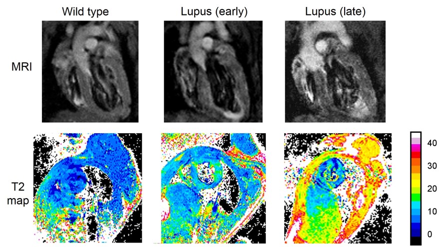
decreased cardiac function in late-stage NZM2410 mice. CMR analysis of sagittal
heart sections showed progressive thickening of the myocardium with a prominent
left ventricular hypertrophy. T2 CMR
mapping showed prolonged T2 signals, consistent with edema, in early and late
stage lupus hearts compared to wild type mice. To examine histopathology,
heart tissue was collected from early and late-stage NZM2410 mice for staining
with H&E and Masson’s trichrome. Evidence of myocarditis and fibrosis was
observed in mice with late-stage disease.

Conclusion: Our results
suggest an association of SLE disease progression with the development of cardiac
abnormalities resulting from inflammation in NZM2410 mice. Therefore, this
animal model could be used to further characterize the pathophysiological
correlation, relationship, and interplay between lupus and cardiovascular
disease in longitudinal studies. Future work will involve identification of
biomarkers related to cardiac involvement and will further develop CMR with
quantitative T2 mapping as a clinical tool to assess inflammation in patients
with SLE.


Disclosure: N. Young, None; J. Hampton, None; S. Aqel, None; J. Pyles, None; A. Bratasz, None; A. Kalyanasundaram, None; W. N. Jarjour, None; S. P. Ardoin, None.

To cite this abstract in AMA style:

Young N, Hampton J, Aqel S, Pyles J, Bratasz A, Kalyanasundaram A, Jarjour WN, Ardoin SP. Progression of Lupus Pathology Is Correlative with Cardiac Magnetic Resonance Imaging Abnormalities, Diminished Function, and Inflammatory Histopathology in an Animal Model [abstract]. Arthritis Rheumatol. 2015; 67 (suppl 10). https://acrabstracts.org/abstract/progression-of-lupus-pathology-is-correlative-with-cardiac-magnetic-resonance-imaging-abnormalities-diminished-function-and-inflammatory-histopathology-in-an-animal-model/. Accessed .
  • Tweet
  • Click to email a link to a friend (Opens in new window) Email
  • Click to print (Opens in new window) Print

« Back to 2015 ACR/ARHP Annual Meeting

ACR Meeting Abstracts - https://acrabstracts.org/abstract/progression-of-lupus-pathology-is-correlative-with-cardiac-magnetic-resonance-imaging-abnormalities-diminished-function-and-inflammatory-histopathology-in-an-animal-model/

Advanced Search

Your Favorites

You can save and print a list of your favorite abstracts during your browser session by clicking the “Favorite” button at the bottom of any abstract. View your favorites »

All abstracts accepted to ACR Convergence are under media embargo once the ACR has notified presenters of their abstract’s acceptance. They may be presented at other meetings or published as manuscripts after this time but should not be discussed in non-scholarly venues or outlets. The following embargo policies are strictly enforced by the ACR.

Accepted abstracts are made available to the public online in advance of the meeting and are published in a special online supplement of our scientific journal, Arthritis & Rheumatology. Information contained in those abstracts may not be released until the abstracts appear online. In an exception to the media embargo, academic institutions, private organizations, and companies with products whose value may be influenced by information contained in an abstract may issue a press release to coincide with the availability of an ACR abstract on the ACR website. However, the ACR continues to require that information that goes beyond that contained in the abstract (e.g., discussion of the abstract done as part of editorial news coverage) is under media embargo until 10:00 AM ET on November 14, 2024. Journalists with access to embargoed information cannot release articles or editorial news coverage before this time. Editorial news coverage is considered original articles/videos developed by employed journalists to report facts, commentary, and subject matter expert quotes in a narrative form using a variety of sources (e.g., research, announcements, press releases, events, etc.).

Violation of this policy may result in the abstract being withdrawn from the meeting and other measures deemed appropriate. Authors are responsible for notifying colleagues, institutions, communications firms, and all other stakeholders related to the development or promotion of the abstract about this policy. If you have questions about the ACR abstract embargo policy, please contact ACR abstracts staff at [email protected].

Wiley

  • Online Journal
  • Privacy Policy
  • Permissions Policies
  • Cookie Preferences

© Copyright 2025 American College of Rheumatology