Session Information
Session Type: ACR Poster Session B
Session Time: 9:00AM-11:00AM
Background/Purpose: Lupus patients are at increased risk for cardiovascular disease (CVD). Chou et al. (Ann Inter Med 2011) found that resting ECG abnormalities, in particular ECG 4-elements (ECG-4), were associated with subsequent CVD events in the general population.
ECG-4 included the following elements: 1) ST-segment and/or T-wave abnormalities; 2) left ventricular hypertrophy (LVH); 3) left axis deviation (LAD) and left anterior fascicular block (LAFB); and 4) left bundle branch block (LBBB) and right bundle branch block (RBBB). However, ECG-4 did not include Q-wave and we have established ECG-5 elements which included Q-wave in addition to the elements of ECG-4.
We aimed to determine the prevalence of ECG-4 and ECG-5 in an SLE cohort and to examine the factors associated with ECG-4 and -5.
Methods: A standard digitally recorded 12-lead resting supine ECG was performed on consecutive patients attending the Lupus Clinic between 2012- 2015. A senior cardiologist interpreted coded ECGs using the Minnesota code classification system. Patients with ECG-4 and -5 were compared to those with normal ECG by t-test and chi-squared test Univariate and multivariate Cox regression models and time-dependent multivariate cox regression models were built to assess the associations between the covariates and ECG-4 and -5.
Descriptive analyses of patients’ demographics, disease activity and damage, and antibodies abnormalities were studied in ECG-4 and -5. For the analysis we used the clinical/laboratory data available at the date of ECG.
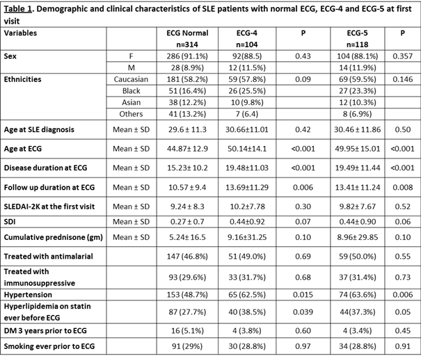
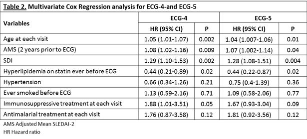
Results: Of 487 patients, 104 (21.4%) and 118 (24.2%) patients had one or more of the ECG-4 and ECG-5 elements respectively. A higher prevalence ECG-4 and -5 was identified among older SLE patients and patients with longer SLE disease duration (Table1). The burden of ECG-4 and ECG-5 elements increased with age. In the univariate analyses, patients with ECG-4 and -5 were older, had a longer lupus disease duration, more active SLE disease (Adjusted Mean SLEDAI-2K 2 years prior to ECG), higher SDI and higher cumulative dose of glucocorticoids compared to patients with normal ECG. The multivariate analyses showed that ECG-4 and -5 were associated with older age and higher SLE disease activity while treatment of hyperlipidemia was protective (Table 2).
Conclusion: We described a novel approach using 5 ECG elements to identify SLE patients at increased risk for CVD events. A high prevalence of ECG-5 (24.2%) was found in this SLE cohort. Controlling SLE disease activity is important since it was associated with ECG-5. Early identification of ECG-5 in SLE patients might allow for better stratification and risk management.
To cite this abstract in AMA style:
Al Rayes H, Harvey P, Gladman DD, Sabapathy A, Urowitz M, Touma Z. Prevalence of ECG Cardiovascular Abnormalities in Lupus Patients: A Novel Approach Using 5 ECG Elements [abstract]. Arthritis Rheumatol. 2016; 68 (suppl 10). https://acrabstracts.org/abstract/prevalence-of-ecg-cardiovascular-abnormalities-in-lupus-patients-a-novel-approach-using-5-ecg-elements/. Accessed .« Back to 2016 ACR/ARHP Annual Meeting
ACR Meeting Abstracts - https://acrabstracts.org/abstract/prevalence-of-ecg-cardiovascular-abnormalities-in-lupus-patients-a-novel-approach-using-5-ecg-elements/


