Session Information
Date: Sunday, October 21, 2018
Title: Systemic Lupus Erythematosus – Etiology and Pathogenesis Poster I
Session Type: ACR Poster Session A
Session Time: 9:00AM-11:00AM
Background/Purpose: Currently, little is known about what distinguishes asymptomatic Anti-Nuclear Antibody (ANA) positive individuals (ANA+NS) who will progress to Systemic Lupus Erythematosus (SLE) from those who will not. Preliminary data from our laboratory indicates that pro-inflammatory cytokines are increased in SLE but not in ANA+ NS. This finding suggests that the development of SLE is characterized by a change in the ability of the autoimmune response to elicit inflammation. Microparticles (MPs) are one of the sources of nuclear antigens in SLE. Previous studies have found increased levels of MP complexed with IgG (MP-ICs) in SLE as compared to healthy controls (HC) and have shown that these MP-ICs constitute a strong pro-inflammatory stimulus. In this study, we examined if ANA+NS individuals have MP-ICs to determine whether the lack of inflammation in these individuals results from the absence, or change in character, of their immune complexes.
Methods: Flow cytometry was used to examine the type, nucleic acid content, and IgG binding of peripheral blood Annexin V+ MPs in ANA–HC (n=7), ANA+NS (≥1:160 by IF, n=11), symptomatic ANA+ individuals lacking sufficient classification criteria for a connective tissue disease diagnosis (UCTD, n=17) and SLE patients (n=10, classified according to the 1997 American College of Rheumatology criteria). MP nucleic acid content was determined by staining with Syto13 which detects DNA and RNA. Eleven specific ANAs were detected using the Bioplex 2200 ANA Screen. The expression levels of five IFN-α induced genes were measured and summed to generate an IFN5 score.
Results:
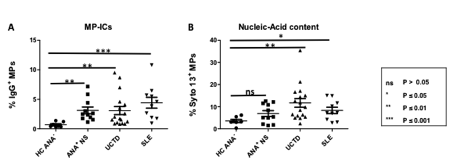
Consistent with previous studies, SLE patients had increased levels of MP-ICs (Figure 1A) that contained higher levels of nucleic acids (Figure 1B), as compared to ANA– HC. Surprisingly, ANA+NS and UCTD patients had similar elevations in the amount of IgG coating their MPs to those seen in SLE (Figure 1A) and in a subset of these individuals the MP nucleic acid content was also higher than ANA– HC (Figure 1B) There was a non-statistically significant trend to higher MP-ICs in ANA+ NS and UCTD individuals with specific ANAs and to higher IFN5 scores in those UCTD individuals with elevated Syto13+ MPs.
Conclusion: MPs appear to be a source of autoantigen in ANA+NS individuals. The results suggest that the differences in elaboration of pro-inflammatory factors between ANA+NS individuals and SLE patients do not result from a lack of immune complexes.
To cite this abstract in AMA style:
Muñoz-Grajales C, Bonilla D, Karanxha A, Ferri D, Silverman E, Johnson S, Bookman A, Touma Z, Wither JE. Presence of Apoptotic Microparticle Containing Immune Complexes in Asymptomatic ANA+ Individuals Despite the Absence of Inflammation [abstract]. Arthritis Rheumatol. 2018; 70 (suppl 9). https://acrabstracts.org/abstract/presence-of-apoptotic-microparticle-containing-immune-complexes-in-asymptomatic-ana-individuals-despite-the-absence-of-inflammation/. Accessed .« Back to 2018 ACR/ARHP Annual Meeting
ACR Meeting Abstracts - https://acrabstracts.org/abstract/presence-of-apoptotic-microparticle-containing-immune-complexes-in-asymptomatic-ana-individuals-despite-the-absence-of-inflammation/

