Session Information
Session Type: ACR Poster Session B
Session Time: 9:00AM-11:00AM
Background/Purpose: In order to visualize amyloid deposition in the specific organs, the patients with systemic amyloidosis underwent positron-emission tomography (PET) study with 11C-BF-227 that specifically binds to aggregated amyloid fibrils. A PET tracer, 11C-BF-227, was previously developed, and we had success with in vivo detection of amyloid plaques in Alzheimerfs disease brains and cardiac amyloidosis in familial transthyretin-related systemic amyloidosis (Kudo Y. et al. in J Nucl Med 8: 2007, and Furukawa K. et al. in Circulation.125:2012, respectively).Here, we investigated additional patients with AA and AL amyloidosis, utilizing this strategy.
Methods: We obtained 6 patients who were diagnosed as amyloidosis with tissue biopsies. We got the informed consent from all study subjects. Patients were 2 female, 4 men (average age plus minus SD, 69 plus minus 5.7) , and they were 2 of Sjogrenfs syndrome, 2 of monoclonal gammopathy of undetermined significance (MGUS), 1 of Rheumatoid Arthritis, and 1 of etiology unknown. The types of amyloid are 3 of AA and 3 of AL amyloid. We also evaluated two healthy controls to compare the images. They were taken 11C-BF-227 PET imaging, then collected an anatomic Magnetic Resonance Image (MRI) sequence to identify the specific organ structures. We analyzed the data with PMOD and Dr. view software, merging the PET and MRI images from the same subject into the same three-dimensional space. The region-of-interest (ROI) was placed on individual MRI images in each organ. The ROI information was then copied onto dynamic PET standardized uptake value (SUV) images. The ratio of regional SUV to the intact lung SUV (SUVR) was also calculated.
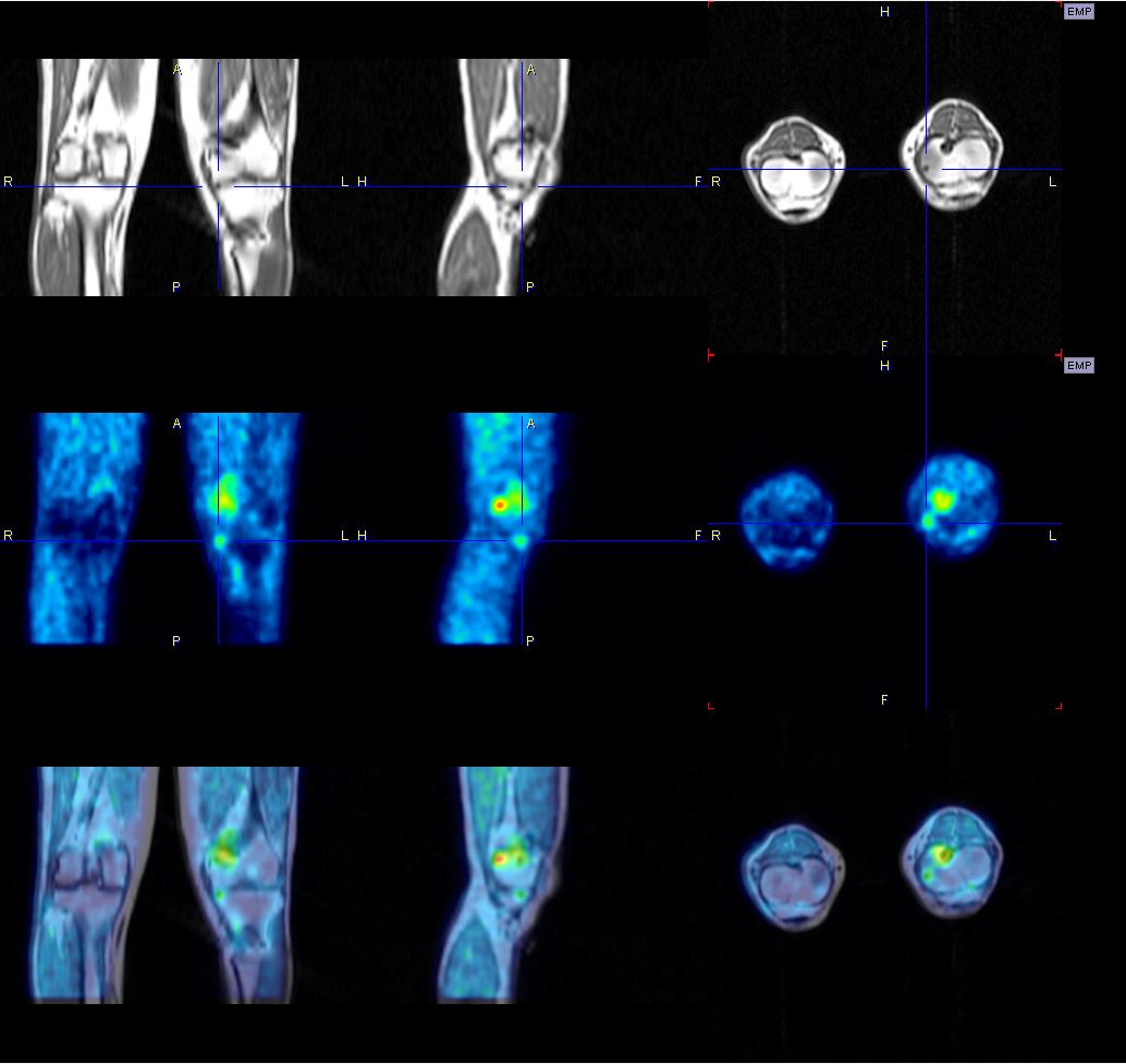
Results: First, we confirmed the amyloid depositions by biopsies of the knee, lung nodules, skin, heart, kidney, and colon. We analyzed the PET images, and the subjects showed the accumulation of 11C-BF-227 in each organ. The representative PET and MRI image of the patient, having amyloid deposition by the knee biopsy, was shown the strong accumulation of 11C-BF-227as indicated in the Figure. His renal and cardiac functions were normal and did not have any amyloid deposition. Another patient having multiple nodules in the lung with amyloid deposition, indicated an increased SUV in those nodules in the PET image. The patients who had amyloid deposition in the kidney showed relatively higher SUVR in the kidney.
Conclusion: We clarified that our newly developed amyloid pet tracer, 11C-BF-227, can detect the amyloid deposition specifically, and 11C-BF-227-PET is a useful technology to detect the amyloid in the whole bodies, although the sensitivity might be different in each organ.
To cite this abstract in AMA style:
Shirota Y, Furukawa K, Tashiro M, Shirrai T, Fujii H, Ishii T, Harigae H. Positron Emission Tomography Images with an Amyloid-Specific Tracer 11C-BF-227 in Systemic Amyloidosis Patients [abstract]. Arthritis Rheumatol. 2016; 68 (suppl 10). https://acrabstracts.org/abstract/positron-emission-tomography-images-with-an-amyloid-specific-tracer-11c-bf-227-in-systemic-amyloidosis-patients/. Accessed .« Back to 2016 ACR/ARHP Annual Meeting
ACR Meeting Abstracts - https://acrabstracts.org/abstract/positron-emission-tomography-images-with-an-amyloid-specific-tracer-11c-bf-227-in-systemic-amyloidosis-patients/

