Session Information
Date: Sunday, November 17, 2024
Title: Vasculitis – Non-ANCA-Associated & Related Disorders Poster II
Session Type: Poster Session B
Session Time: 10:30AM-12:30PM
Background/Purpose: Ultrasonography can measure vascular intimal-medial thickness (IMT) and delineate the degrees of stenosis in Takayasu arteritis (TAK). Further, higher IMT has been reported in patients with active disease, with prospective studies showing changes in carotid IMT with treatment. In this study we measured IMT at multiple vascular sites at enrollment and at six months and assessed the association between the IMT and clinical characteristics, inflammatory markers and PET-VAS scores. We also tried to correlate change in IMT with change in disease activity, hsCRP, serum pentraxin-3, serum amyloid-A, serum IgG.
Methods: We prospectively enrolled 49 TAK patients (fulfilling ACR 1993) from May 2022 to December 2023; 25 had active and 24 inactive disease state as per Physician Global assessment (PGA). Arterial IMT was measured at 8 vascular sites (bilateral common carotid, subclavian, brachial, and femoral arteries) using ultrasonography (Esaote MyLab50) by two different observers. Observers were blinded between each other and towards the clinical details. For each vascular site, mean of three highest values obtained from fixed segments was obtained. Thickness was measured in the far wall with uniform frequency, angle of insonation and depth. Disease activity was evaluated using Indian Takayasu Arteritis Activity Score (ITAS 2010), inflammatory markers and PET VAS score. PGA was considered the gold standard measure for classifying active and inactive disease. Patients were followed-up and their arterial IMT and disease activity parameters were reassessed at 6 months.
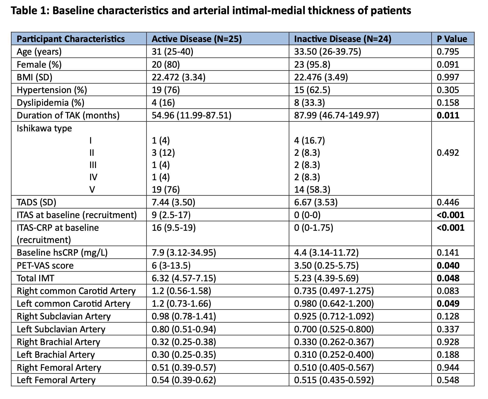
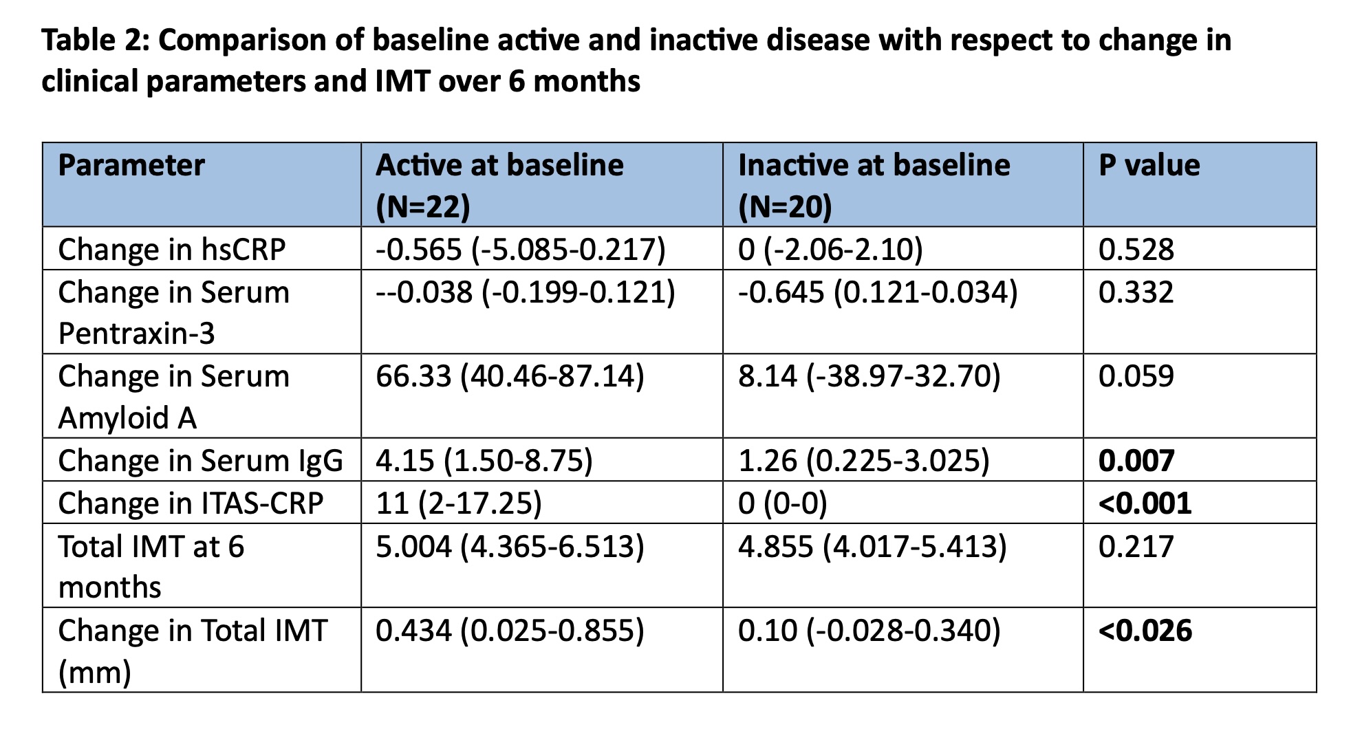
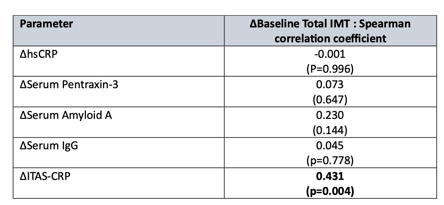
Results: Most patients(68.08%) had an Ishikawa type V disease. Interobserver agreement was adequate. Total IMT of 8 arteries and that of left common carotid artery were significantly higher in the active group (table 1). Forty two patients were reassessed at 6-months follow-up. Mean total IMT was significantly lower at 6-months(median 4.93 mm) compared to baseline(median 5.23mm) with p value < 0.001. Change in total IMT correlated positively with a change in disease activity status as measured by change in ITAS-CRP. No significant correlations were observed between change in carotid IMT and changes in inflammatory markers(table 2). Change in IMT over 6 months was significantly higher in patients who had active disease at recruitment.
Conclusion: Ultrasonographic measurement of IMT at multiple arterial sites, particularly carotid arteries, is a promising tool for evaluating TAK disease activity. Measurement of total carotid IMT alone may be a sufficient marker for disease activity in supradiaphragmatic disease. There is significant change in IMT over 6 months especially in the group with active disease, suggesting that multivessel IMT is a potential biomarker for disease modification in Takayasu arteritis which is simple and non-invasive.
To cite this abstract in AMA style:
Jose A, Thabah M, Kavadichanda C, K L J, Mariaselvam C, Negi V. Multi-vessel Intimal Medial Thickness in Takayasu Arteritis: A Potential Marker for Disease Modification? [abstract]. Arthritis Rheumatol. 2024; 76 (suppl 9). https://acrabstracts.org/abstract/multi-vessel-intimal-medial-thickness-in-takayasu-arteritis-a-potential-marker-for-disease-modification/. Accessed .« Back to ACR Convergence 2024
ACR Meeting Abstracts - https://acrabstracts.org/abstract/multi-vessel-intimal-medial-thickness-in-takayasu-arteritis-a-potential-marker-for-disease-modification/


