Session Information
Session Type: Poster Session (Tuesday)
Session Time: 9:00AM-11:00AM
Background/Purpose: Osteoarthritis (OA) is a degenerative joint disease characterized by deterioration of articular cartilage in the joints. MRI T1ρ and T2 relaxation times have been proposed as potential biomarkers for early detection of cartilage degeneration. However, few studies examined their reliability in a multi-site multi-vendor setting, which is critical for future large-scale trials and clinical translation of such quantitative measures. The purpose of this study was to evaluate the intra-site repeatability and inter-site reproducibility of knee cartilage T1ρ and T2 data acquired by multiple sites and multiple vendors in phantoms and human subjects.
Methods: Four 3T MR systems from four different sites and three vendors (Siemens, GE, and Philips) with dedicated knee coils were used for this study (Table 1a). Five subjects were scanned with same day rescan at each site, including two traveling volunteers whose knees were scanned at all four sites. The imaging protocol included 3D T1ρ and T2 imaging, high-resolution gradient echo (GRE) imaging or dual echo steady state (DESS), and 3D turbo spin echo imaging (Table 1b). Data were transferred to one site for centralized data post-processing using in-house developed software. For all relaxation time fitting, two-parameter mono-exponential fittings were performed. The echo time or spin lock times used for fitting are listed in Table 1c. Cartilage was segmented semi-automatically into six compartments (medial/lateral femur [MFC/LFC], medial/lateral tibia [MT/LT, trochlea [TRO], and patellar [PAT]) using high-resolution DESS/GRE images, and the segmentation was overlaid on T1ρ and T2 maps after registration to calculate means and standard deviations of T1ρ and T2 values in each compartment.
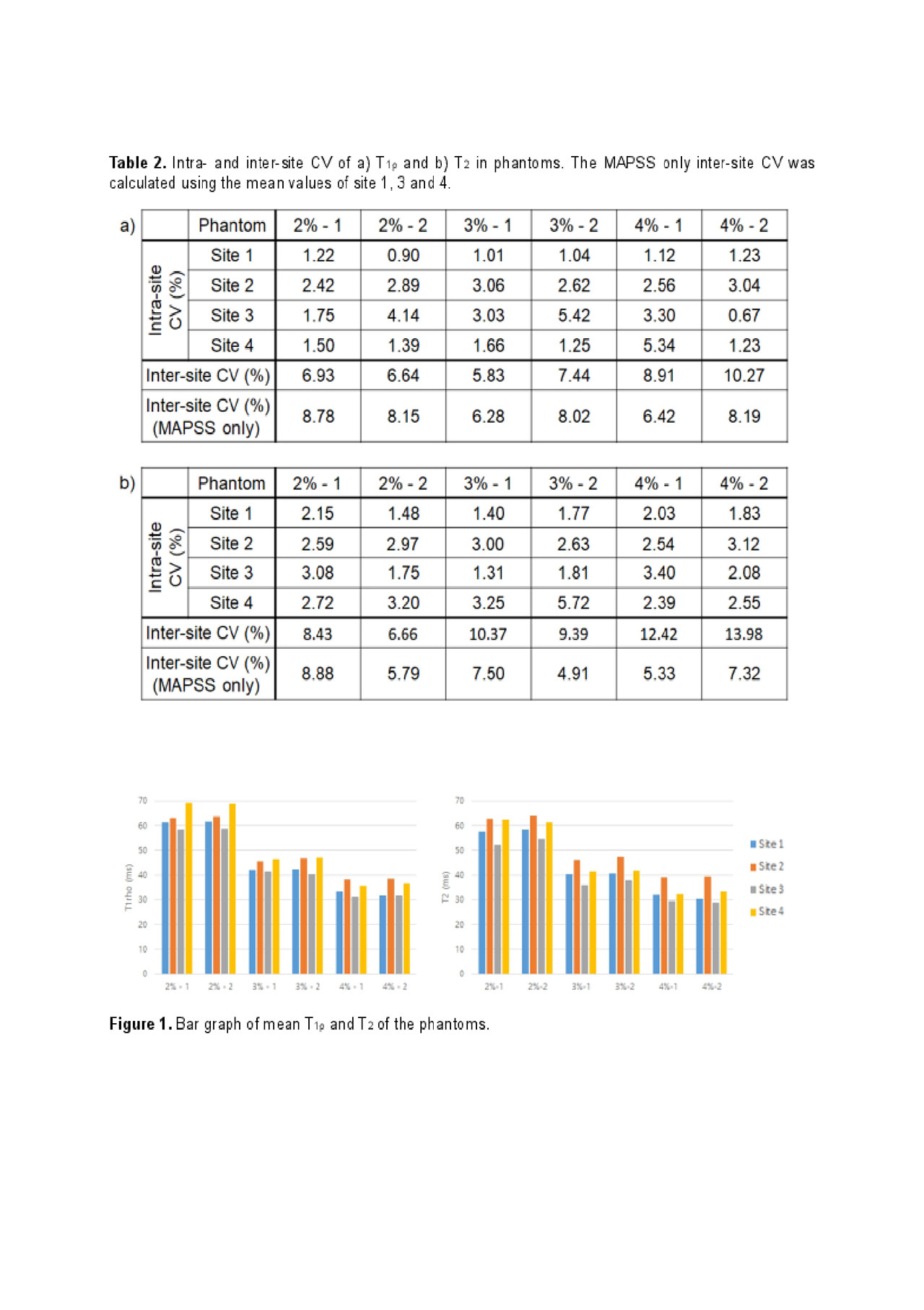
Results: For phantoms, all sites showed excellent intra-site repeatability of T1ρ and T2 values with an average CV of 2.24% for T1ρ and of 2.53% for T2 (Table 2, Fig. 1). The average inter-site CVs were 7.67 % and 10.21 % for T1ρ and T2 respectively with both sequences included (four sites); and 7.64% and 6.62% for T1ρ and T2 respectively with the MAPSS sequence only (three sites).
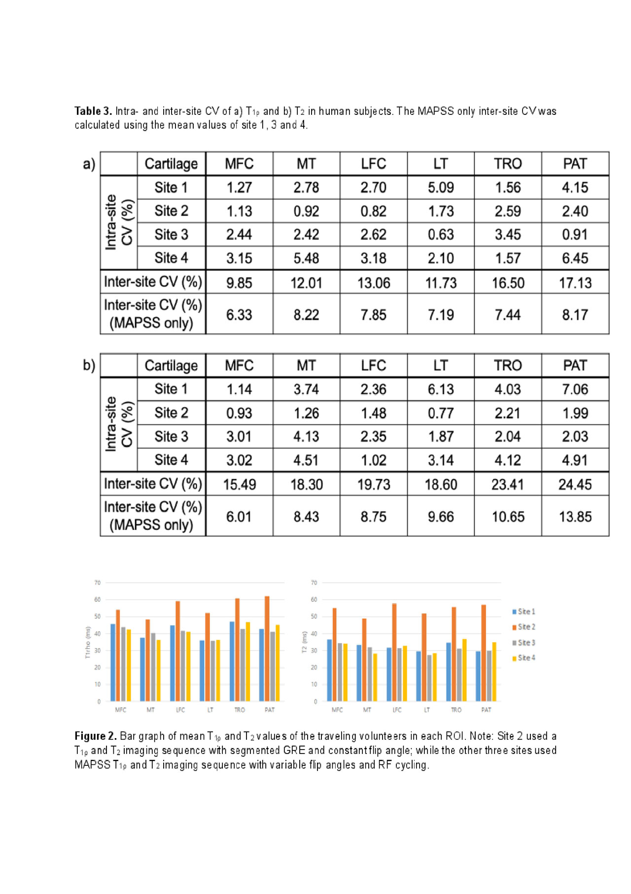
For human subjects, excellent intra-site repeatability was observed for all sites with an average CV of 2.33% for T1ρ and of 2.91% for T2, Table 3. The average inter-site CVs were 13.50% and 28.15% for T1ρ and T2 respectively with both sequences included (four sites); and 8.2% and 10.17% for T1ρ and T2 respectively with MAPSS sequences only (three sites). Fig. 2 shows the T1ρ and T2 values of each defined cartilage compartment for the traveling volunteers.
Conclusion: Cartilage T1ρ and T2 imaging, after standardization of data acquisition and post-processing, demonstrates excellent intra-site repeatability, and inter-site reproducibility, suggesting great promise that T1ρ and T2 may serve as reliable imaging biomarkers for future multi-site and multi-vendor studies, after standardization of data acquisition and post-processing. Factors that could introduce variations in T1ρ and T2 values such as temperature, B0 and B1 inhomogeneity will be investigated further in future studies.
To cite this abstract in AMA style:
Kim J, Mamoto K, Lartey R, Xu K, Tanaka M, Bahroos E, Winalski C, Link T, Hardy P, Peng Q, Botto-van Bemden A, Liu K, Peters R, Wu C, Li X. Multi-vendor Multi-site T1ρ and T2 Quantification of Knee Cartilage [abstract]. Arthritis Rheumatol. 2019; 71 (suppl 10). https://acrabstracts.org/abstract/multi-vendor-multi-site-t1%cf%81-and-t2-quantification-of-knee-cartilage/. Accessed .« Back to 2019 ACR/ARP Annual Meeting
ACR Meeting Abstracts - https://acrabstracts.org/abstract/multi-vendor-multi-site-t1%cf%81-and-t2-quantification-of-knee-cartilage/


