Session Information
Session Type: ACR Abstract Session
Session Time: 2:30PM-4:00PM
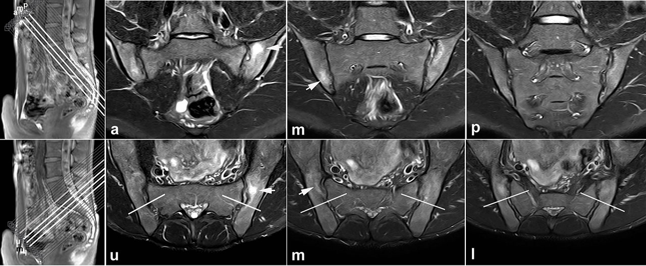
Background/Purpose: Low grade bone marrow edema (BME) on magnetic resonance imaging (MRI) of the sacroiliac joints (SIJ) is challenging the discrimination between patients with early axial spondyloarthritis (SpA) and mechanical back pain. In our previous analysis, recreational and elite athletes had on average 3-4 SIJ quadrants with BME, and 30-41% met the Assessment of SpondyloArthritis international Society (ASAS) definition of active sacroiliitis [1]. Potential conditions simulating SIJ BME such as vascular partial volume effect or anatomical SIJ variants could not be explored due to standard semi-coronal MRI scans only. Our goals by assessing combined semi-axial and semi-coronal SIJ MRI scans in 2 cohorts of young athletes were to explore the frequency and topography of non-specific BME by 2 perpendicular MRI planes, its association with 4 constitutional SIJ features, and potential limitation of false positive assignments of ASAS-defined sacroiliitis by standard semi-coronal scans alone.
Methods: Combined semi-axial and semi-coronal SIJ MRI scans of 20 recreational runners before/after running and 22 elite ice-hockey players were evaluated by 3 blinded readers for BME and its association with 4 constitutional SIJ features: vascular partial volume effect, deep iliac ligament insertion, fluid-filled bone cyst, and lumbosacral transitional anomaly. Scans of TNF-treated SpA patients served to mask readers. Pre-test reader agreement for BME on semi-axial scans was calculated by single measure, absolute agreement intra-class correlation coefficient (ICC). We analysed distribution and topography of BME and associated constitutional SIJ features across 8 anatomical SIJ regions (upper/lower ilium/sacrum, subdivided in anterior/posterior slices) descriptively, as concordantly recorded by ≥2/3 readers on both MRI planes. BME confirmed on both scans was compared with previous evaluation of semi-coronal MRI alone which met the ASAS definition for active sacroiliitis.
Results: ICC agreement among 3 readers for semi-axial SIJ BME was 0.86 (0.72-0.95). Perpendicular semi-axial and semi-coronal MRI scans confirmed SIJ BME consistently in 25% and 27% of athletes, preferentially in the anterior upper sacrum. BME associated with 4 constitutional SIJ features was observed in 20-36% of athletes, clustering in the posterior lower ilium. The proportion of ASAS-positive sacroiliitis recorded on semi-coronal plane alone decreased by 30-50% upon amending semi-axial scans (from 30-35% to 20% in runners, from 41% to 18% in ice-hockey players).
Conclusion: Semi-axial combined with standard semi-coronal scans in MRI protocols for sacroiliitis facilitated recognition of non-specific BME, which clustered in the posterior lower ilium in association with constitutional SIJ features, and in the anterior upper sacrum. The proportion of false-positive ASAS assignments of sacroiliitis recorded on semi-coronal plane alone could be substantially reduced by amending semi-axial scans.
Reference. [1] Weber U et al. Arthritis Rheum 2018;70:736.
To cite this abstract in AMA style:
Weber U, Jurik A, Zejden A, Larsen E, Jørgensen S, Rufibach K, Schioldan C, Schmidt-Olsen S. MRI of the Sacroiliac Joints in Athletes: Do Semi-axial Slices Added to Standard Semi-coronal Scans Facilitate Recognition of Non-specific Bone Marrow Edema? [abstract]. Arthritis Rheumatol. 2019; 71 (suppl 10). https://acrabstracts.org/abstract/mri-of-the-sacroiliac-joints-in-athletes-do-semi-axial-slices-added-to-standard-semi-coronal-scans-facilitate-recognition-of-non-specific-bone-marrow-edema/. Accessed .« Back to 2019 ACR/ARP Annual Meeting
ACR Meeting Abstracts - https://acrabstracts.org/abstract/mri-of-the-sacroiliac-joints-in-athletes-do-semi-axial-slices-added-to-standard-semi-coronal-scans-facilitate-recognition-of-non-specific-bone-marrow-edema/


