Session Information
Session Type: Abstract Submissions (ACR)
· Background/Purpose: Change in joint space narrowing (JSN) on radiographs is currently the gold standard for monitoring disease progression of knee osteoarthritis in clinical trials and is considered a surrogate marker for cartilage volume. However, this method can be unreliable due to high measurement error and it is insensitive to early change. Furthermore, cross-sectional studies have shown that JSN not only reflects hyaline articular cartilage but also meniscal pathology. Therefore, the aim of this study was to quantitate the correlation between changes in meniscal extrusion and cartilage volume loss over two years and radiographic osteoarthritis (ROA) change over ten years.
· Methods: A sample of 220 subjects (mean age 45 years at baseline; age range 26–61 years) was evaluated at baseline, two, and ten years. Approximately half were adult offspring of subjects who had a knee replacement performed for knee osteoarthritis and the remaining were randomly selected controls that were initially matched by age and sex. Knee cartilage volume and meniscal extrusion were determined using T1-weighted fat-suppression MRI techniques at baseline and two years. ROA, JSN, and osteophytes were scored using fixed flexion radiographs at baseline and ten year using standard protocols. Spearman-ranked correlation analysis was used to analyse the correlation between changes in medial meniscal extrusion (MME) and medial tibial cartilage (MTC) loss with changes in ROA, JSN, and osteophytes. All were adjusted for age, sex and BMI.
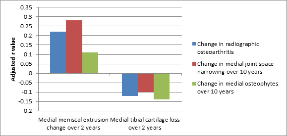
· Results: The mean MTC volume was 2234mm3 at baseline with a 2.5% loss per annum over two years. At baseline, 8% of participants had any medial meniscal extrusion and 3% had an incident meniscal extrusion over two years. An increase in MME over two years predicted a change in ROA over ten years in adjusted analysis (r=0.22, p=0.003), an increase in medial JSN (r=0.30, p=<0.001) and an increase in medial osteophytes (r=0.15, p=0.046) [Fig 1]. However, a change in MTC over two years did not correlate with a change in ROA over ten years (r=-0.12, p=0.096) or a change in medial JSN (r=-0.10, p=0.182) and weakly with change in medial osteophytes (r=-0.14, p=0.045) [Fig 1].
· Conclusion: In a midlife cohort at higher risk of osteoarthritis, a change in MME, despite being quite rare, is more strongly correlated with subsequent change in ROA than cartilage volume loss. These findings suggest ROA is a composite measure of joint pathology and does not primarily or sufficiently reflect cartilage loss.
Figure 1. Correlation between changes in medial meniscal extrusion and medial tibial cartilage loss over two years with radiographic changes over ten years
Disclosure:
L. Chou,
None;
H. I. Khan,
None;
A. McBride,
None;
D. Aitken,
None;
C. Ding,
None;
L. Blizzard,
None;
J. P. Pelletier,
ArthroLab,
4;
J. martel-Pelletier,
ArthroLab,
4;
F. Cicuttini,
None;
G. Jones,
None.
« Back to 2013 ACR/ARHP Annual Meeting
ACR Meeting Abstracts - https://acrabstracts.org/abstract/meniscal-extrusion-is-a-better-predictor-of-radiographic-osteoarthritis-change-over-ten-years-compared-to-cartilage-volume-loss/

