ACR Meeting Abstracts

ACR Meeting Abstracts

  • Meetings
    • ACR Convergence 2024
    • ACR Convergence 2023
    • 2023 ACR/ARP PRSYM
    • ACR Convergence 2022
    • ACR Convergence 2021
    • ACR Convergence 2020
    • 2020 ACR/ARP PRSYM
    • 2019 ACR/ARP Annual Meeting
    • 2018-2009 Meetings
    • Download Abstracts
  • Keyword Index
  • Advanced Search
  • Your Favorites
    • Favorites
    • Login
    • View and print all favorites
    • Clear all your favorites
  • ACR Meetings

Abstract Number: 1809

Melanocytes Are Driven Toward an Antigen Presentation Phenotype Through UV-Induced Keratinocyte Crosstalk and Exposure to Type I Interferons in Patients with Cutaneous Lupus Erythematosus

Rezvan Moallemian, Lin Zhang, Mehrnaz Gharaee-Kermani, Allison Billi, Amy Hurst, Benjamin Klein and J. Michelle Kahlenberg, University of Michigan, Ann Arbor, MI

Meeting: ACR Convergence 2024

Keywords: Cutaneous, Systemic lupus erythematosus (SLE)

  • Tweet
  • Click to email a link to a friend (Opens in new window) Email
  • Click to print (Opens in new window) Print
Session Information

Date: Monday, November 18, 2024

Title: SLE – Etiology & Pathogenesis Poster

Session Type: Poster Session C

Session Time: 10:30AM-12:30PM

Background/Purpose: Cutaneous lupus erythematosus (CLE) encompasses various lesion morphologies, but photosensitivity and high type I interferon (IFN) responses are a unifying theme amongst all CLE subtypes. Chronic, elevated type I IFN exposure heightens inflammatory responses of keratinocytes (KCs) to ultraviolet radiation (UV), leading to additional type I IFN production and exacerbation of lesions. Interactions between keratinocytes and other skin cells, including melanocytes, play a crucial role in the regulation of inflammatory responses and protection against stressors.

Methods: We utilized single-cell RNA sequencing (scRNA-seq) to analyze gene expression profiles in melanocyte populations from skin samples from systemic lupus erythematosus (SLE) patients and healthy controls with and without UV treatment. Primary melanocytes were isolated from healthy control skin and stimulated with type I and type II interferons to observe their response via bulk RNA-sequencing and real-time qPCR. Flow cytometry was utilized to further characterize the crosstalk between melanocytes and keratinocytes. This was done by exposing keratinocytes to UV radiation and subsequently incubating primary melanocytes with the supernatant from these keratinocytes.

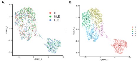
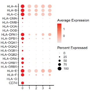
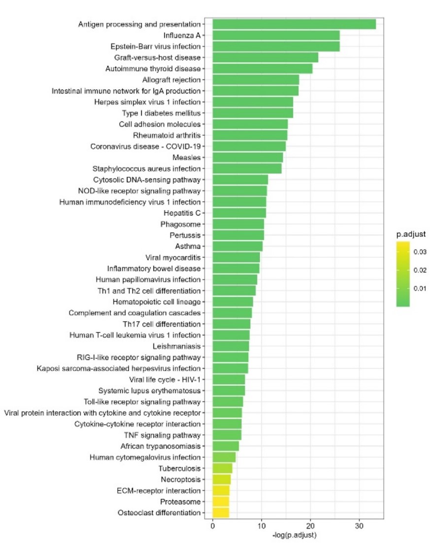
Results: We identified an unusual population of melanocytes in lesional and non-lesional SLE skin that is characterized by high type I IFN-regulated gene expression and upregulation of MHC class II (Figures 1 and 2). Bulk RNAseq data from primary melanocytes stimulated with type I and II interferons showed that treatment with either IFNα, IFNβ and or IFNγ was sufficient to increase antigen presentation genes in melanocytes (Figure 3). We thus hypothesized that KC-derived IFNs generated in response to UV light may be modulating melanocyte function. Indeed, we found that supernatants from UV-treated keratinocytes induce upregulation of antigen-presenting markers in melanocytes and this is increased when KCs are exposed to a high type I IFN environment, as is present in SLE skin. Ongoing experiments are evaluating the KC-derived factors that induce these changes in melanocytes and are examining the antigen presenting function of melanocytes.

Conclusion: Melanocytes isolated from SLE skin are abnormal and are characterized by IFN exposure and MHC Class II upregulation. Our data suggests type I IFNs induce MHC Class II upregulation in melanocytes and may be shifting melanocyte function from keratinocyte protection to immune activation. Future research aims to elucidate melanocyte-mediated antigen presentation and T cell activation in SLE.

Supporting image 1

Figure 1. UMAPS of melanocyte populations from 14 healthy control (H) and seven paired non-lesional and lesional skin biopsies from SLE patients shown by (A) disease status or (B) cluster. SLE Melanocytes exhibit unique transcriptional profiles in non-lesional (NLE) and lesional (LE) skin that identify antigen presentation capacity on MHC class II.

Supporting image 2

Figure 2. Dot plot of antigen presentation-related transcripts that are seen across all clusters. Cluster 0 is composed entirely of non-lesional and lesional samples and identifies significant antigen presentation capacity on MHC class I and II, compared with all the other clusters.

Supporting image 3

Figure 3. KEGG pathway analyses of the 334 commonly up-regulated genes from IFNα / IFNβ / IFNγ vs control. Antigen processing and presentation is the dominant downstream pathway following IFNα, β and γ stimulation in primary melanocytes. Primary melanocytes from healthy controls were stimulated with IFNα, β and γ for 24 hours. RNA was then isolated and subjected to bulk RNA sequencing. Adjusted p-value cutoff: 0.05 ; q-value cutoff: 0.2


Disclosures: R. Moallemian: None; L. Zhang: None; M. Gharaee-Kermani: None; A. Billi: None; A. Hurst: None; B. Klein: None; J. Kahlenberg: Amgen, 12, coauthor on publication, AstraZeneca, 2, Bristol-Myers Squibb(BMS), 2, 5, Eli Lilly, 2, Gilead, 2, GlaxoSmithKlein(GSK), 2, Janssen, 5.

To cite this abstract in AMA style:

Moallemian R, Zhang L, Gharaee-Kermani M, Billi A, Hurst A, Klein B, Kahlenberg J. Melanocytes Are Driven Toward an Antigen Presentation Phenotype Through UV-Induced Keratinocyte Crosstalk and Exposure to Type I Interferons in Patients with Cutaneous Lupus Erythematosus [abstract]. Arthritis Rheumatol. 2024; 76 (suppl 9). https://acrabstracts.org/abstract/melanocytes-are-driven-toward-an-antigen-presentation-phenotype-through-uv-induced-keratinocyte-crosstalk-and-exposure-to-type-i-interferons-in-patients-with-cutaneous-lupus-erythematosus/. Accessed .
  • Tweet
  • Click to email a link to a friend (Opens in new window) Email
  • Click to print (Opens in new window) Print

« Back to ACR Convergence 2024

ACR Meeting Abstracts - https://acrabstracts.org/abstract/melanocytes-are-driven-toward-an-antigen-presentation-phenotype-through-uv-induced-keratinocyte-crosstalk-and-exposure-to-type-i-interferons-in-patients-with-cutaneous-lupus-erythematosus/

Advanced Search

Your Favorites

You can save and print a list of your favorite abstracts during your browser session by clicking the “Favorite” button at the bottom of any abstract. View your favorites »

All abstracts accepted to ACR Convergence are under media embargo once the ACR has notified presenters of their abstract’s acceptance. They may be presented at other meetings or published as manuscripts after this time but should not be discussed in non-scholarly venues or outlets. The following embargo policies are strictly enforced by the ACR.

Accepted abstracts are made available to the public online in advance of the meeting and are published in a special online supplement of our scientific journal, Arthritis & Rheumatology. Information contained in those abstracts may not be released until the abstracts appear online. In an exception to the media embargo, academic institutions, private organizations, and companies with products whose value may be influenced by information contained in an abstract may issue a press release to coincide with the availability of an ACR abstract on the ACR website. However, the ACR continues to require that information that goes beyond that contained in the abstract (e.g., discussion of the abstract done as part of editorial news coverage) is under media embargo until 10:00 AM ET on November 14, 2024. Journalists with access to embargoed information cannot release articles or editorial news coverage before this time. Editorial news coverage is considered original articles/videos developed by employed journalists to report facts, commentary, and subject matter expert quotes in a narrative form using a variety of sources (e.g., research, announcements, press releases, events, etc.).

Violation of this policy may result in the abstract being withdrawn from the meeting and other measures deemed appropriate. Authors are responsible for notifying colleagues, institutions, communications firms, and all other stakeholders related to the development or promotion of the abstract about this policy. If you have questions about the ACR abstract embargo policy, please contact ACR abstracts staff at [email protected].

Wiley

  • Online Journal
  • Privacy Policy
  • Permissions Policies
  • Cookie Preferences

© Copyright 2025 American College of Rheumatology