Session Information
Date: Tuesday, November 14, 2023
Title: (2387–2424) Vasculitis – Non-ANCA-Associated & Related Disorders Poster III
Session Type: Poster Session C
Session Time: 9:00AM-11:00AM
Background/Purpose: Magnetic resonance imaging (MRI) is well established for diagnosing giant cell arteritis (GCA). Its role in monitoring disease activity has yet to be determined. We investigated vascular and musculoskeletal inflammation using MRI in the patients of the GUSTO trial.
Methods: Eighteen patients with newly diagnosed GCA received 500 mg methylprednisolone intravenously for 3 consecutive days [1]. After that, GC treatment was discontinued, and a single dose of tocilizumab (TCZ) was administered intravenously, followed by weekly subcutaneous TCZ injections from day 10 until week 52. Cranial, thoracic and abdominal MR exams were performed at baseline (active, new-onset disease), and at weeks 24, 52 (remission on-treatment), and 104 (remission off-treatment). Polymyalgia rheumatica (PMR) findings as well as vasculitic disease extent and intensity were rated by experienced radiologistsand one board-certified neuroradiologist as previously reported (grade 0-3, grade ≥ 2 considered as vasculitis) [2,3]. Up to 10 cranial, 13 large vessel and 18 musculoskeletal segments were assessed.
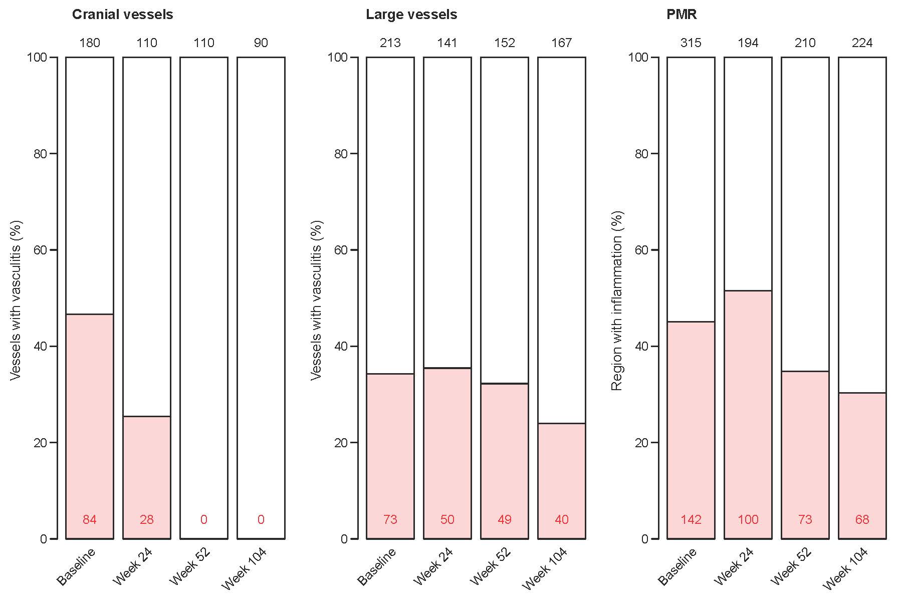
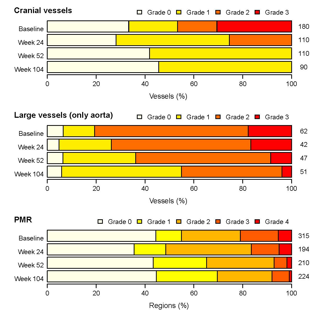
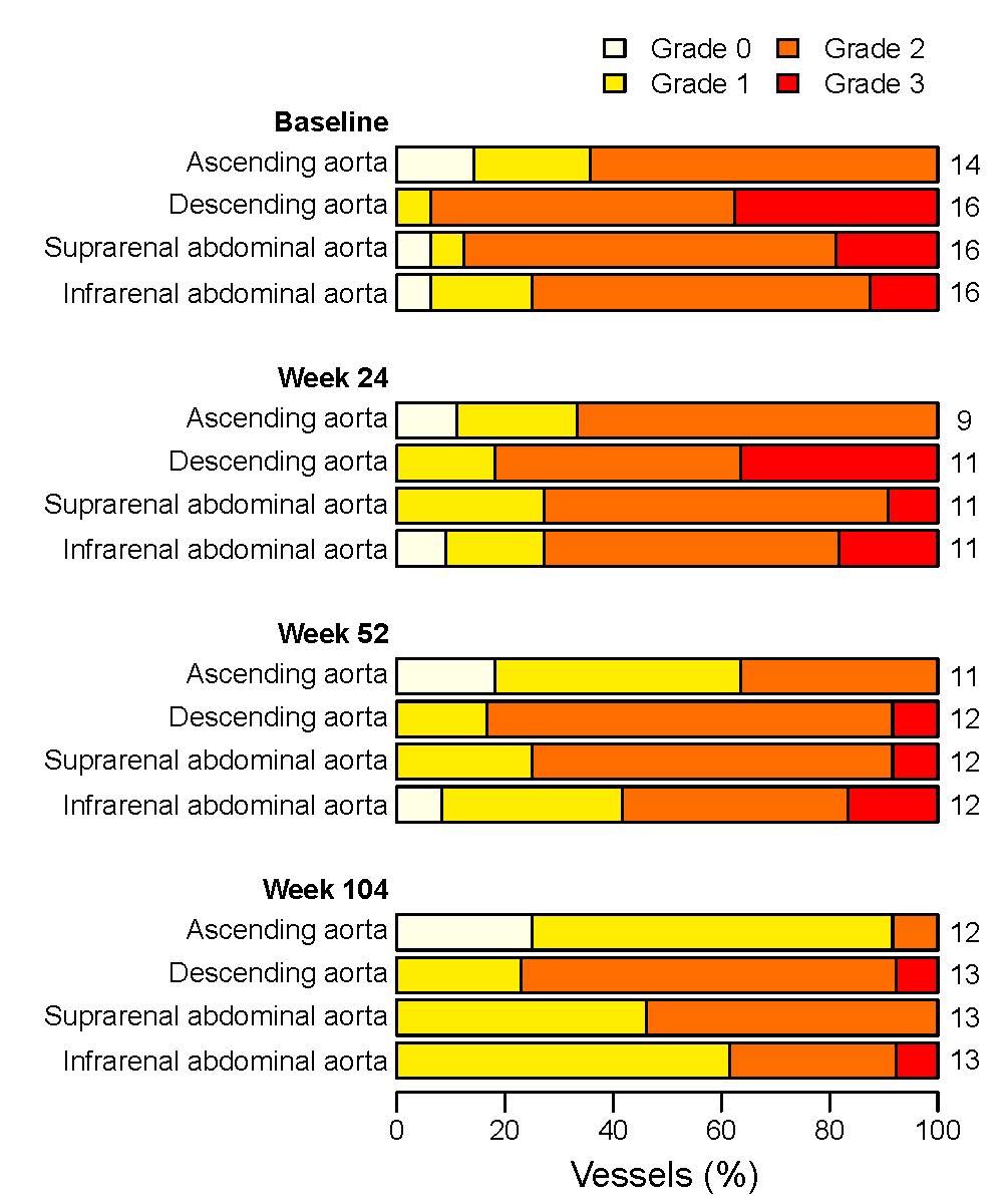
Results: In total, 673 vascular segments and 943 musculoskeletal regions in 56 thoracic/abdominal MR and 490 vascular segments in 49 cranial MR scans of 18 patients were analyzed. Cranial vessels displayed dynamic changes. At weeks 52 and 104, no cranial vascular segment displayed vasculitic findings. Vasculitic signal alterations were still detectable in every 4th cranial segment at week 24. Large vessels, except for the ascending aorta, displayed no or only a slight decrease in vasculitic findings upon remission on- and off-treatment. PMR findings persisted in most patients. Results are displayed in Figures 1 – 3.
Conclusion: Whereas signals of cranial vessels normalized upon a 52-week treatment, large vessel signals and PMR findings persisted. Remission of vasculitic activity in cranial arteries took more than 24 weeks, which was longer than time to clinical symptom resolution and normalization of serum proteins but paralleled intima-media thickness measurements using ultrasound [4]. Thus, in contrast to large vessel signals and PMR findings, the dynamic of cranial vessel signals suggests that MRI of these arteries is a very promising monitoring tool for suspected relapse after 52 weeks of treatment. References: 1. Christ et al. Lancet Rheumatology, 2021
2. Klink et al. Radiology, 2014.
3. Reichenbach et al. Rheumatology, 2018.
4. Seitz et al. Rheumatology, 2021.
To cite this abstract in AMA style:
Christ L, Bonel H, Cullmann J, Seitz L, Butikofer L, Wagner F, Villiger P. Magnetic Resonance Imaging Reflects Disease Activity in the “Giant Cell Arteritis Treatment with Ultra-short Glucocorticoids and Tocilizumab” Trial (The GUSTO Trial) [abstract]. Arthritis Rheumatol. 2023; 75 (suppl 9). https://acrabstracts.org/abstract/magnetic-resonance-imaging-reflects-disease-activity-in-the-giant-cell-arteritis-treatment-with-ultra-short-glucocorticoids-and-tocilizumab-trial-the-gusto-trial/. Accessed .« Back to ACR Convergence 2023
ACR Meeting Abstracts - https://acrabstracts.org/abstract/magnetic-resonance-imaging-reflects-disease-activity-in-the-giant-cell-arteritis-treatment-with-ultra-short-glucocorticoids-and-tocilizumab-trial-the-gusto-trial/


