Session Information
Date: Sunday, November 10, 2019
Title: Innate Immunity Poster
Session Type: Poster Session (Sunday)
Session Time: 9:00AM-11:00AM
Background/Purpose: Salt, or sodium, has recently emerged as a potent factor involved in modulating inflammatory responses. Sodium is implicated as a dietary risk factor for (ACPA-positive) RA1 and increased sodium concentrations lead to enhanced Th17 cell differentiation2. Furthermore, sodium accumulates locally in certain inflamed and sclerotic tissues such as the skin, both in the context of infection and in autoimmunity, as has been shown in scleroderma patients3. To clarify whether sodium, as a possible inflammation-enhancing factor, may also accumulate locally in the joints in autoimmune arthritis, we used a state-of-the-art, non-invasive 7-Tesla 23Na MRI technique to measure sodium concentrations in inflamed knees of patients with autoimmune joint disease.
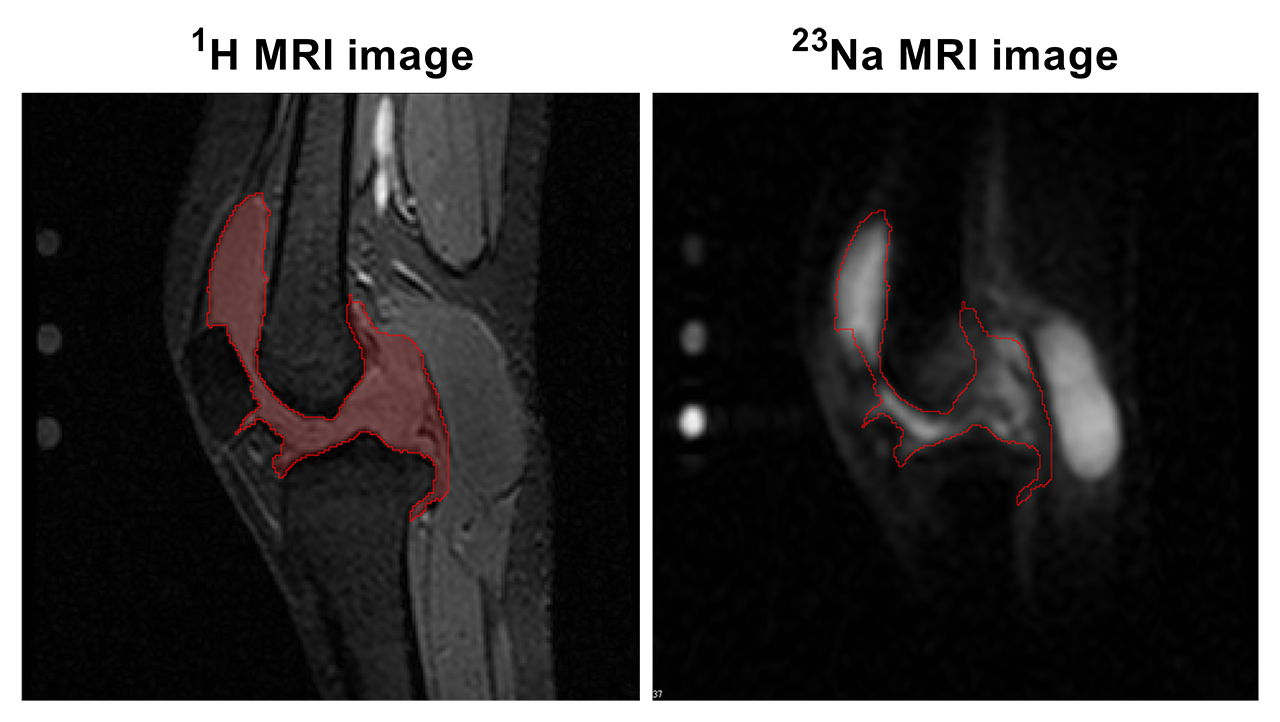
Methods: In 8 patients with autoimmune arthritis of the knee (3 JIA, 2 PsA, 2 RA, 1 peripheral SpA) and 11 healthy controls, we measured synovial Na+ content in the (affected) knee with a birdcage double-tuned proton/23Na sodium transmit/receive coil on a 7-Tesla MR system (Philips Healthcare, Best, the Netherlands). Cartilage, synovium, and synovial fluid (in the intra-articular space and synovial bursae) was manually segmented in the proton image in six sagittal slices, after which the volume of interest was transferred to the 23Na image for extraction of 23Na MR signal intensity. Average Na+ content in mmol/L was calculated by relating 23Na MR signal intensity to three agarose standard gels of known Na+ concentration (20, 50, and 100 mmol/L), scanned in tandem with each knee (see Figure 1).
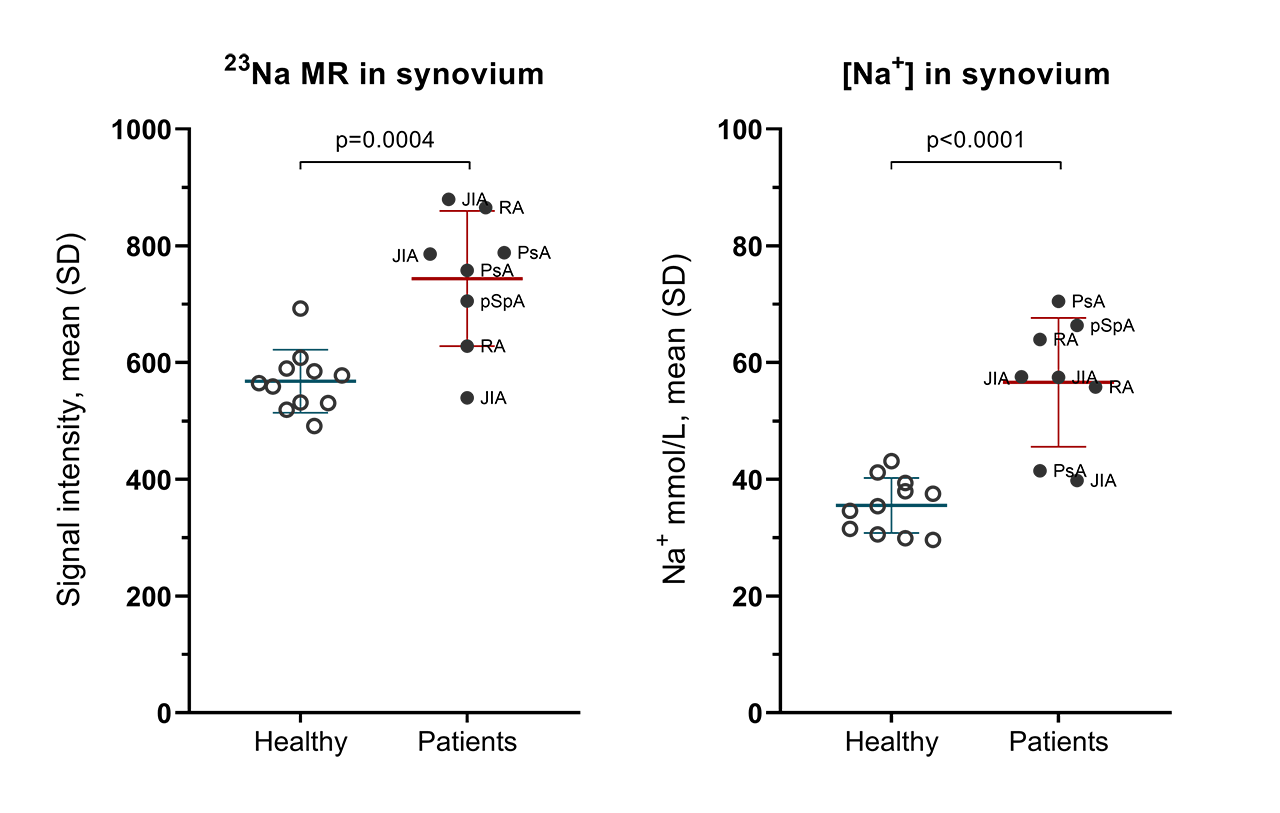
Results: 23Na-MRI signal intensity in synovial structures was consistent and reproducible across slices: between slice coefficient of variation in a representative patient was 9.4%. 23Na-MRI demonstrated increased 23Na MR signal intensity in arthritis knees of patients compared to healthy controls: mean (SD): 744 (116) vs 568 (54), p=0.0004 (Figure 2). When this MR signal intensity was converted to Na+ concentration, patients had clearly increased Na+-concentrations compared to controls: mean (SD): 57 (11) vs 36 (5) mmol/L, p< 0.0001. A linear regression with synovial Na+ concentration as the outcome indicated that the differences in synovial Na+ concentration between patients and healthy controls could not be explained by differences in synovial volume (not shown).
Conclusion: Tissue sodium detection and quantification by 7-Tesla 23Na MRI is feasible and reliable, and indicates significantly higher sodium content in inflamed knees of patients with autoimmune joint disease compared to healthy controls. This suggests that sodium may be involved in modulating inflammation and synovial cell biology. Whether this is specific for chronic autoimmune joint diseases or may be present for more general inflammation remains to be determined.
References:
- Jiang et al. 2016 ARD: doi:10.1136/annrheumdis-2015-209009
- Wu et al. 2013 Nature: doi:10.1038/nature11984
- Kopp et al. 2017 Rheumatology: doi:10.1093/rheumatology/kew371
To cite this abstract in AMA style:
de Moel E, Teeuwisse W, Huizinga T, Toes R, Webb A, van der Woude D. Increased Sodium Accumulation Detected by 23Na-Magnetic Resonance Imaging in Inflamed Knees of Patients with Autoimmune Joint Disease [abstract]. Arthritis Rheumatol. 2019; 71 (suppl 10). https://acrabstracts.org/abstract/increased-sodium-accumulation-detected-by-23na-magnetic-resonance-imaging-in-inflamed-knees-of-patients-with-autoimmune-joint-disease/. Accessed .« Back to 2019 ACR/ARP Annual Meeting
ACR Meeting Abstracts - https://acrabstracts.org/abstract/increased-sodium-accumulation-detected-by-23na-magnetic-resonance-imaging-in-inflamed-knees-of-patients-with-autoimmune-joint-disease/


