Session Information
Date: Monday, November 14, 2016
Title: Cytokines, Mediators, Cell-Cell Adhesion, Cell Trafficking and Angiogenesis - Poster I
Session Type: ACR Poster Session B
Session Time: 9:00AM-11:00AM
Background/Purpose: Cutaneous lupus erythematosus (CLE) is an autoimmune disease with various subsets and wide-ranging clinical manifestations. T lymphocytes are the predominant cell type found in lesions, but plasmacytoid dendritic cells (pDCs) and myeloid dendritic cells (mDCs) also play a pivotal role in the pathogenesis. Although antimalarials are the primary treatment for CLE, the exact mode of action is still not completely understood. To figure out the determining factor of treatment response to antimalarials in CLE patients, inflammatory cell infiltrates and cytokine profiles in lesional skin of CLE patients were evaluated.
Methods: Thirty-two skin biopsies of CLE patients (SCLE; n=13, DLE; n=19) were immunohistochemically investigated for the presence of pDCs, mDCs, neutrophils, and macrophages by employing antibodies against CD123, CD11c, MPO, and MAC387 respectively. Skin sections were examined by light microscopy and positive stained cells were quantified at x400 magnification in five non-overlapping adjacent microscopic fields in the dermis. The results were expressed as mean number of cells. RNA extracted from formalin-fixed paraffin-embedded (FFPE) skin samples from 21 CLE patients were analyzed by qRT-PCR for gene expression of type I IFN signature genes (LY6E, OAS1, OASL, ISG15, and MX1) and inflammatory cytokines TNFα. The patients were grouped according to their response to antimalarial treatment; 1) Hydroxychloroquine (HCQ) group, who were responsive to HCQ; and 2) HCQ + Quinacrine (QC) group, who were refractory to HCQ and responded to additional QC. The Mann-Whitney test was used to compare the number of each cell type and gene expression between HCQ and QC responsive groups.
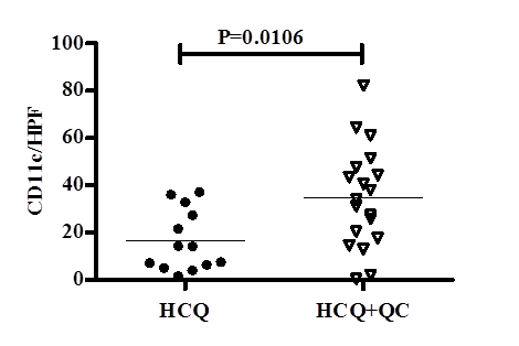
Results: The number of mDCs was significantly higher (p=0.0106) in HCQ+QC group patients (19 cases) compared to those in HCQ group (13 cases) (Figure). The mean number (standard error) of pDCs, mDCs, neutrophils, and macrophages in each group of patients is summarized in the table. Gene expression of type I IFN signatures were significantly upregulated in the HCQ group compared to the HCQ+QC group (LYE p=0.0037; OAS1 p=0.0002; OASL p=0.0067; ISG15 p=0.01; and MX1 p=0.0083), while TNFα levels were significantly higher in the HCQ+QC group (p=0.003).
Conclusion: Increased numbers of mDCs together with higher TNFα expression might be responsible for refractoriness to HCQ and better response to QC. Our data suggest that the increased number of mDCs in CLE patients may be used as a predictive factor of refractoriness to HCQ and responsiveness to QC.
| Table | ||||||||||||||||||
|
*SE: Standard Error; HCQ: Hydroxychloroquine group; HCQ+QC: Hydroxychloroquine + Quinacrine group
Figure
To cite this abstract in AMA style:
Zeidi M, Kim HJ, Werth VP. Increased Population of Myeloid Dendritic Cells and Upregulated Gene Expression of Tnfα Is Associated with Poor Response to Hydroxychloroquine [abstract]. Arthritis Rheumatol. 2016; 68 (suppl 10). https://acrabstracts.org/abstract/increased-population-of-myeloid-dendritic-cells-and-upregulated-gene-expression-of-tnf%ce%b1-is-associated-with-poor-response-to-hydroxychloroquine/. Accessed .« Back to 2016 ACR/ARHP Annual Meeting
ACR Meeting Abstracts - https://acrabstracts.org/abstract/increased-population-of-myeloid-dendritic-cells-and-upregulated-gene-expression-of-tnf%ce%b1-is-associated-with-poor-response-to-hydroxychloroquine/

