Session Information
Session Type: Poster Session A
Session Time: 10:30AM-12:30PM
Background/Purpose: Growth arrest specific protein 6 (Gas6) is a member of the vitamin K-dependent protein family. It participates in apoptosis, inflammatory response and immunomodulation by binding to its receptors Tyro3, Axl and Mer (TAM). Previous studies have suggested that Gas6 is able to affect the coagulation system by regulating platelet and vascular cell function through the TAM signaling pathway. However, its role in antiphospholipid syndrome (APS) is unclear. This research focuses on the expression level and clinical significance of Gas6 in APS to further understand the role of Gas6/TAM signaling pathway in the pathogenesis of APS.
Methods: 102 patients diagnosed APS with Sydney criteria were recruited from the Department of Rheumatology and Immunology at Peking University People’s Hospital. 104 healthy volunteers were enrolled as control. Among them, the serum Gas6 concentrations of 92 APS patients were determined by ELISA, and the relationship between Gas6 and clinical characteristics was analyzed. The expression of TAM receptor on the surface of peripheral blood monocytes in 10 APS patients was further examined. The results were analyzed by Mann-Whitney U test, Kruskal-Wallis test followed by Dunn’s posttest for multiple comparisons and Spearman’s rank correlation test as appropriate. p < 0. 05 was considered significant.
Results: Compared to HC, APS patients had higher serum expression of Gas6 (11.81±5.02 vs. 9.22±3.22 ng/mL, p < 0.0001) (Fig.1A). Furthermore, serum Gas6 concentrations in patients with thrombotic APS (tAPS) and obstetric APS (oAPS) were analyzed. While oAPS was higher than tAPS (12.21 ± 4.057 vs. 11.42 ± 5.537 ng/mL, p = 0.0476) (Fig.1B). And serum Gas6 concentrations in tAPS (p = 0.0203) and oAPS (p < 0.0001) were significantly higher than in HC. In PAPS (11.63 ± 4.655 ng/mL, p < 0.0001) and SAPS (12.28 ± 5.973 ng/mL, p = 0.0008), serum Gas6 concentrations were higher than in HC, with no significant difference between PAPS and SAPS (p > 0.9999) (Fig. 1C). Next, the correlation between patients’ serum Gas6 concentrations and clinical features was explored. The results showed that patients’ serum Gas6 concentrations showed negative correlation with HGB (r = −0.4581,p < 0.0001), C3 (r = −0.4026,p = 0.0023), and APTT (r = −0.3657,p = 0.0172), and positive correlation with IgG (r = 0.3819,p = 0.0024) and ESR (r = 0.4982,p =0.0032) (Fig.2). Moreover, Gas6 concentration was higher in patients with low C3 (p = 0.0015) and low C4 (p < 0.0001) than in patients with normal complement, and were also higher in ANA-positive patients than in ANA-negative patients (p = 0.0101). The expression level of Tyro3 on the surface of peripheral blood monocytes in APS (MFI: 11.01 ± 14.44) was significantly higher than HC (MFI: 1.744 ± 0.7233, p = 0.0015) (Fig. 3B). For Axl (MFI: 1.136 ± 0.2480 vs. 0.9975 ± 0.2477, p = 0.2176) and Mer (MFI: 4.022 ± 2.257 vs. 3.576 ± 1.049, p = 0.9705), no significant differences were detected between APS and HC (Fig. 3C/D).
Conclusion: Serum Gas6 was significantly overexpressed in APS patients and correlated with parameters of inflammation, immunity and coagulation, suggesting that the Gas6/TAM signaling pathway may play a potential role in APS.
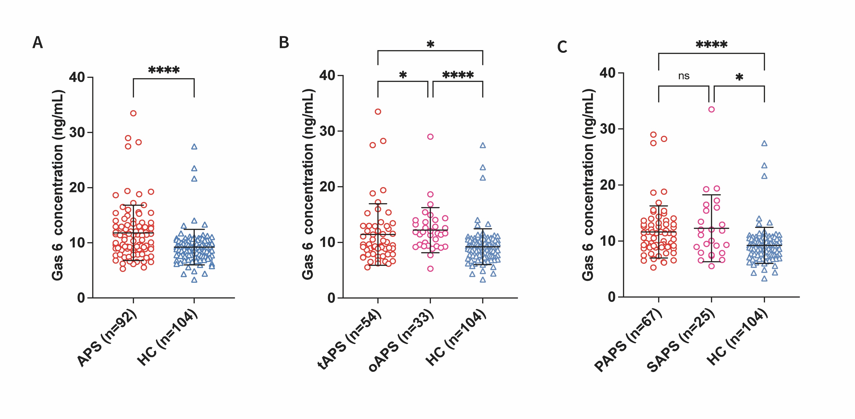 Fig.1. A) Serum Gas6 concentration was significantly higher in antiphospholipid syndrome (APS) patients than in healthy controls (HC) (p < 0.0001). B) Serum Gas6 concentration was significantly higher in both thrombotic antiphospholipid syndrome (tAPS) (p = 0.0203) and obstetric antiphospholipid syndrome (oAPS) (p < 0.0001) patients, and the concentration in the oAPS was significantly higher (p = 0.0476). C) Serum Gas6 concentration was significantly higher in both primary antiphospholipid syndrome (PAPS) (p < 0.0001) and secondary antiphospholipid syndrome (SAPS) (p = 0.0008) patients, and there was no significant difference between them (p > 0.9999).
.jpg) Fig.2. Correlation analysis between serum Gas6 concentration and clinical features in APS patients, which showed negative correlation with HGB (A), C3 (B), APTT (E), and positive correlation with IgG (C) and ESR (D).
Fig.2. Correlation analysis between serum Gas6 concentration and clinical features in APS patients, which showed negative correlation with HGB (A), C3 (B), APTT (E), and positive correlation with IgG (C) and ESR (D).
.jpg) Fig.3. A) Monocytes in the peripheral blood of APS patients were sorted by CD14 and CD16. B) The expression level of Tyro3 on the surface of monocytes in the peripheral blood of APS was significantly higher than HC (p = 0.0015). C/D) There was no significant difference in the expression levels of Axl (p = 0.2176) and Mer (p = 0.9705) between APS and HC.
Fig.3. A) Monocytes in the peripheral blood of APS patients were sorted by CD14 and CD16. B) The expression level of Tyro3 on the surface of monocytes in the peripheral blood of APS was significantly higher than HC (p = 0.0015). C/D) There was no significant difference in the expression levels of Axl (p = 0.2176) and Mer (p = 0.9705) between APS and HC.
To cite this abstract in AMA style:
Li F, Yao R, Hu F, Xu L, Li C. Increased Expression of Gas6 and Its Tyrosine Kinase Receptor Tyro3 Are Associated with Antiphospholipid Syndrome [abstract]. Arthritis Rheumatol. 2025; 77 (suppl 9). https://acrabstracts.org/abstract/increased-expression-of-gas6-and-its-tyrosine-kinase-receptor-tyro3-are-associated-with-antiphospholipid-syndrome/. Accessed .« Back to ACR Convergence 2025
ACR Meeting Abstracts - https://acrabstracts.org/abstract/increased-expression-of-gas6-and-its-tyrosine-kinase-receptor-tyro3-are-associated-with-antiphospholipid-syndrome/