ACR Meeting Abstracts

ACR Meeting Abstracts

  • Meetings
    • ACR Convergence 2024
    • ACR Convergence 2023
    • 2023 ACR/ARP PRSYM
    • ACR Convergence 2022
    • ACR Convergence 2021
    • ACR Convergence 2020
    • 2020 ACR/ARP PRSYM
    • 2019 ACR/ARP Annual Meeting
    • 2018-2009 Meetings
    • Download Abstracts
  • Keyword Index
  • Advanced Search
  • Your Favorites
    • Favorites
    • Login
    • View and print all favorites
    • Clear all your favorites
  • ACR Meetings

Abstract Number: 1719

High-Throughput Quantitative Histology in Systemic Sclerosis Skin Disease Using Computer Vision

Chase Correia1, Seamus Mawe2, Mary A. Carns3, Kathleen Aren3, Aileen Hoffman4, Monique Hinchcliff5 and J. Matthew Mahoney6, 1Northwestern University, Chicago, IL, 2Department of Neurological Sciences, University of Vermont Larner College of Medicine, Burlington, VT, 3Northwestern University, Feinberg School of Medicine Scleroderma Program, Chicago, IL, 4Northwestern Feinberg School of Medicine, Chicago, IL, 5Rheumatology, Northwestern University, Chicago, IL, 6Neurological Sciences, University of Vermont College of Medicine, Burlington, VT

Meeting: 2018 ACR/ARHP Annual Meeting

Keywords: histopathologic, skin fibrosis and systemic sclerosis

  • Tweet
  • Click to email a link to a friend (Opens in new window) Email
  • Click to print (Opens in new window) Print
Session Information

Date: Monday, October 22, 2018

Title: Systemic Sclerosis and Related Disorders – Clinical Poster II

Session Type: ACR Poster Session B

Session Time: 9:00AM-11:00AM

Background/Purpose: The lack of a reproducible and quantitative method to accurately assess histological skin fibrosis in patients with systemic sclerosis (SSc) has long plagued clinical studies. Deep neural network (DNN) analysis, a computer vision technique, has the potential to radically augment histopathological analyses for myriad diseases including SSc. DNN analysis is a computational tool that extracts and quantifies complex image patterns. To test the hypothesis that DNN analysis applied to SSc skin biopsies is a useful method for dermal fibrosis quantification, the association between DNN outputs and skin score was determined.

Methods: One rheumatologist performed modified Rodnan skin score (mRSS) assessments followed by four mm dermal, punch biopsies of the non-dominant volar forearm, in patients with SSc. Biopsies were repeated at 6-, 12- 24-, and 36- months. Trichrome-stained sections were photomicrographed and transformed into quantitative features using AlexNet in the Matlab Neural Network Toolbox. Principal component analysis was used to identify the combination of quantitative features that captured the most quantitative feature variance. The correlation between the first principal component, mRSS, and local arm skin score was assessed.

Results: Twenty-six photomicrographs were analyzed. DNN analysis identified 4096 unique quantitative features. The first principal component of these 4096 quantitative features strongly correlated with local skin score (R = 0.52) and mRSS (R = 0.71).

Conclusion: This investigation demonstrates that DNN-derived quantitative features of SSc biopsies are sensitive to both local skin score and mRSS. Future directions include testing for generalizability in a larger sample and evaluating whether DNN are sensitive to biochemical measures of fibrosis. If validated, this suggests that DNN analysis can dramatically expand the quantifiable SSc phenome to permit inclusion of histological information into SSc trajectory models.
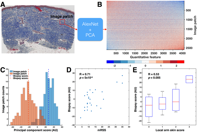
 

Figure 1:

A) 100 image patches (red square) were randomly sampled from the dermis and input to AlexNet. B) For each image patch, AlexNet identified 4096 quantitative features, resulting in a 2600 x 4096-dimensional matrix. The rows and columns of the quantitative feature matrix have been sorted by the first principal component (PC). C) Histograms of the principal component scores for image patches from the patients with the highest (35) and lowest (4) mRSS show little overlap. Across all 2600 image patches grouped by biopsy, the biopsy accounted for 30% of the total variance in the principal component scores (p < 10-100, one-way ANOVA). Therefore, biopsies were summarized into biopsy scores by taking the mean of the 100 patches. D) Biopsy score significantly correlated with mRSS (Pearson’s R = 0.71, p = 5×10⁻⁵). E) Biopsy score significantly correlated to local skin score (Pearson’s R = 0.53, p = 0.0049).


Disclosure: C. Correia, None; S. Mawe, None; M. A. Carns, None; K. Aren, None; A. Hoffman, None; M. Hinchcliff, None; J. M. Mahoney, None.

To cite this abstract in AMA style:

Correia C, Mawe S, Carns MA, Aren K, Hoffman A, Hinchcliff M, Mahoney JM. High-Throughput Quantitative Histology in Systemic Sclerosis Skin Disease Using Computer Vision [abstract]. Arthritis Rheumatol. 2018; 70 (suppl 9). https://acrabstracts.org/abstract/high-throughput-quantitative-histology-in-systemic-sclerosis-skin-disease-using-computer-vision/. Accessed .
  • Tweet
  • Click to email a link to a friend (Opens in new window) Email
  • Click to print (Opens in new window) Print

« Back to 2018 ACR/ARHP Annual Meeting

ACR Meeting Abstracts - https://acrabstracts.org/abstract/high-throughput-quantitative-histology-in-systemic-sclerosis-skin-disease-using-computer-vision/

Advanced Search

Your Favorites

You can save and print a list of your favorite abstracts during your browser session by clicking the “Favorite” button at the bottom of any abstract. View your favorites »

All abstracts accepted to ACR Convergence are under media embargo once the ACR has notified presenters of their abstract’s acceptance. They may be presented at other meetings or published as manuscripts after this time but should not be discussed in non-scholarly venues or outlets. The following embargo policies are strictly enforced by the ACR.

Accepted abstracts are made available to the public online in advance of the meeting and are published in a special online supplement of our scientific journal, Arthritis & Rheumatology. Information contained in those abstracts may not be released until the abstracts appear online. In an exception to the media embargo, academic institutions, private organizations, and companies with products whose value may be influenced by information contained in an abstract may issue a press release to coincide with the availability of an ACR abstract on the ACR website. However, the ACR continues to require that information that goes beyond that contained in the abstract (e.g., discussion of the abstract done as part of editorial news coverage) is under media embargo until 10:00 AM ET on November 14, 2024. Journalists with access to embargoed information cannot release articles or editorial news coverage before this time. Editorial news coverage is considered original articles/videos developed by employed journalists to report facts, commentary, and subject matter expert quotes in a narrative form using a variety of sources (e.g., research, announcements, press releases, events, etc.).

Violation of this policy may result in the abstract being withdrawn from the meeting and other measures deemed appropriate. Authors are responsible for notifying colleagues, institutions, communications firms, and all other stakeholders related to the development or promotion of the abstract about this policy. If you have questions about the ACR abstract embargo policy, please contact ACR abstracts staff at [email protected].

Wiley

  • Online Journal
  • Privacy Policy
  • Permissions Policies
  • Cookie Preferences

© Copyright 2025 American College of Rheumatology