ACR Meeting Abstracts

ACR Meeting Abstracts

  • Meetings
    • ACR Convergence 2024
    • ACR Convergence 2023
    • 2023 ACR/ARP PRSYM
    • ACR Convergence 2022
    • ACR Convergence 2021
    • ACR Convergence 2020
    • 2020 ACR/ARP PRSYM
    • 2019 ACR/ARP Annual Meeting
    • 2018-2009 Meetings
    • Download Abstracts
  • Keyword Index
  • Advanced Search
  • Your Favorites
    • Favorites
    • Login
    • View and print all favorites
    • Clear all your favorites
  • ACR Meetings

Abstract Number: 2921

High Resolution 3D Fast Spin-Echo T1 Black-Blood Imaging for the Diagnosis of Giant Cell Arteritis

Christine rodriguez 1, Wagih Ben Hassen 1, Pierre Seners 2, Catherine Oppenheim 1 and Alexis Régent3, 1Département d’imagerie, pôle Neuro Sainte Anne, GHT Paris – Psychiatrie & Neurosciences, 1 rue Cabanis, 75014 Paris, Paris, Ile-de-France, France, 2Service de neurologie, pôle Neuro Sainte Anne, GHT Paris – Psychiatrie & Neurosciences, 1 rue Cabanis, 75014 Paris, Paris, Ile-de-France, France, 3National Referral Center for Rare Systemic Autoimmune Diseases Paris Cochin, Paris, France

Meeting: 2019 ACR/ARP Annual Meeting

Keywords: giant cell arteritis, magnetic resonance imaging (MRI) and diagnostic imaging

  • Tweet
  • Email
  • Print
Session Information

Date: Wednesday, November 13, 2019

Title: 6W024: Vasculitis – Non-ANCA-Associated & Related Disorders III: Large Vessel Vasculitis Pathogenesis & Imaging (2918–2923)

Session Type: ACR Abstract Session

Session Time: 11:00AM-12:30PM

Background/Purpose: Giant cell arteritis (GCA) is the most common form of vasculitis of large arteries affecting people older than 50 years. Temporal artery biopsy (TAB) is still considered as the gold standard for GCA diagnosis but it is invasive and its sensitivity ranges from 39 to 86%.Color Doppler Ultrasonography and PET-scanner have been proposed as alternative methods for GCA diagnosis but are not widespread in routine clinical practice. Recent recommendations suggest the use of MRI as an alternative method for GCA diagnosis. To our knowledge, there is no evaluation of 3D sequences in the diagnosis of GCA

Methods: We decided to evaluate the diagnostic performance of pre and post-contrast fat-suppressed 3D-Fast Spin echo T1-weighted black-blood magnetic resonance imaging (CUBE-T1), of the external carotid in the diagnosis process of GCA.

Results: Thirty-two patients (7 men, 25 women, mean±SD age 70.2±12.1 years) were included. The final diagnosis, blinded to CUBE T1 results, was GCA for 10 of them. 3D CUBE T1 displayed the arteries wall clearly, allowing an easy identification of parietal enhancement and the 3D TOF coregistration was helpful in image analysis. Intra- and inter-observer agreement for mural enhancement on CUBE T1 was 1 and 0.84, respectively.

Height of the 10 patients with GCA had a strong mural enhancement in CUBE-T1 (Figure 1), while none of the 22 patients without GCA had mural enhancement in MR images. Therefore, sensitivity was 80% and specificity was 100 %. The positive predictive value of post-contrast CUBE T1 was 100% and the negative predictive value was 92%. In our cohort, only 6 of the 10 GCA patients had a positive TAB. Therefore sensitivity of TAB was 60%. If TAB was used as the reference standard, MRI yielded a sensitivity of 100%. Two patients with a final diagnosis of GCA despite normal TAB had an arteritis on MRI and vessel enhancement was observed in occipital artery only for one patient.

Conclusion: We here demonstrate the feasibility and accuracy of a 3D post-contrast CUBE-T1 in the diagnosis of GCA. Indeed, we observed a sensitivity of 80%, which is higher than the 64.1% that was observed by Rhéaume et al. with 2D sequences on his series of 172 patients. We hypothesize that 3D MRI acquisition allowing multiplanar reconstructions together with the 3D TOF coregistration might explain the higher sensitivity and the excellent reproducibility. The reproducibility and short scan duration is a clear advantage of CUBE-T1 sequences with sub-millimetric voxels acquisition. Additional studies are required to confirm our preliminary results.
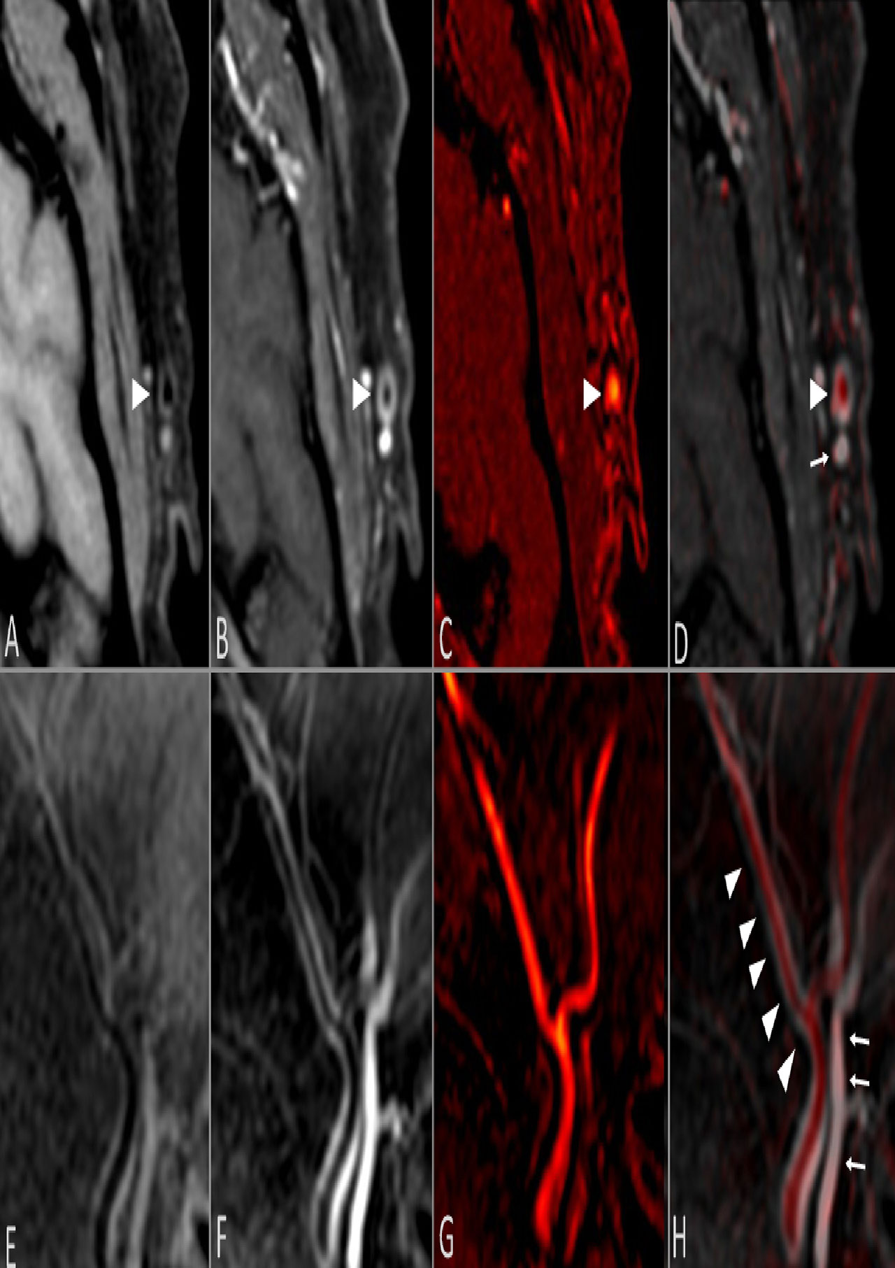

Figure 1

FIGURE 1. Mural enhancement of the left temporal artery in giant cell arteritis.
Axial and sagittal reconstructions of CUBE T1 sequence before -A and E respectively- and after contrast injection -B and F respectively-. 3D TOF sequence in axial and sagittal plane -C and G respectively- coregistered with post-contrast CUBE T1 sequence -D and H respectively-, allowing a differentiation between the temporal artery in red -arrowhead- -C, G- and the temporal vein -arrows- -D, H-. Strong mural enhancement of the temporal artery -arrowhead- -B, D, F, H-.


Disclosure: C. rodriguez, None; W. Ben Hassen, None; P. Seners, None; C. Oppenheim, None; A. Régent, None.

To cite this abstract in AMA style:

rodriguez C, Ben Hassen W, Seners P, Oppenheim C, Régent A. High Resolution 3D Fast Spin-Echo T1 Black-Blood Imaging for the Diagnosis of Giant Cell Arteritis [abstract]. Arthritis Rheumatol. 2019; 71 (suppl 10). https://acrabstracts.org/abstract/high-resolution-3d-fast-spin-echo-t1-black-blood-imaging-for-the-diagnosis-of-giant-cell-arteritis/. Accessed .
  • Tweet
  • Email
  • Print

« Back to 2019 ACR/ARP Annual Meeting

ACR Meeting Abstracts - https://acrabstracts.org/abstract/high-resolution-3d-fast-spin-echo-t1-black-blood-imaging-for-the-diagnosis-of-giant-cell-arteritis/

Advanced Search

Your Favorites

You can save and print a list of your favorite abstracts during your browser session by clicking the “Favorite” button at the bottom of any abstract. View your favorites »

All abstracts accepted to ACR Convergence are under media embargo once the ACR has notified presenters of their abstract’s acceptance. They may be presented at other meetings or published as manuscripts after this time but should not be discussed in non-scholarly venues or outlets. The following embargo policies are strictly enforced by the ACR.

Accepted abstracts are made available to the public online in advance of the meeting and are published in a special online supplement of our scientific journal, Arthritis & Rheumatology. Information contained in those abstracts may not be released until the abstracts appear online. In an exception to the media embargo, academic institutions, private organizations, and companies with products whose value may be influenced by information contained in an abstract may issue a press release to coincide with the availability of an ACR abstract on the ACR website. However, the ACR continues to require that information that goes beyond that contained in the abstract (e.g., discussion of the abstract done as part of editorial news coverage) is under media embargo until 10:00 AM ET on November 14, 2024. Journalists with access to embargoed information cannot release articles or editorial news coverage before this time. Editorial news coverage is considered original articles/videos developed by employed journalists to report facts, commentary, and subject matter expert quotes in a narrative form using a variety of sources (e.g., research, announcements, press releases, events, etc.).

Violation of this policy may result in the abstract being withdrawn from the meeting and other measures deemed appropriate. Authors are responsible for notifying colleagues, institutions, communications firms, and all other stakeholders related to the development or promotion of the abstract about this policy. If you have questions about the ACR abstract embargo policy, please contact ACR abstracts staff at [email protected].

Wiley

  • Online Journal
  • Privacy Policy
  • Permissions Policies
  • Cookie Preferences

© Copyright 2025 American College of Rheumatology