Session Information
Session Type: Abstract Submissions (ACR)
Background/Purpose
Rheumatoid arthritic (RA) disease progression and anti-rheumatic treatment efficacy have traditionally been monitored by evaluation of radiographs. However, MRI has emerged as a more sensitive tool to depict synovial and articular inflammatory changes in early and low disease activity that are not radiographically evident. In particular, T1ρ MRI allows the detection of early-stage cartilage damage1. There are no prior reports of using T1ρ to evaluate cartilage composition changes in the wrist of RA patients. In this study we assessed the feasibility of wrist cartilage T1ρ mapping in RA patients and the association of T1ρ values with RA MRI scoring (RAMRIS).
Methods
The wrists of 5 RA patients (55.2 ± 13.3 yrs.,4 fem., DASCRP28: 3.72 ± 2.26, RA ) and two healthy subjects (27 ± 4 yrs., 1 fem.) were scanned on a 3T MR scanner (GE, Healthcare) using the standard imaging protocol recommended by the OMERACT group and a coronal T1ρ (time of spin-lock = 0/10/20/50 ms; spin-lock frequency = 500Hz, resolution 0.21×0.21×3 mm)2. For both volunteers, scan/re-scan data were acquired after repositioning. Radiocarpal joint cartilage was segmented semi-automatically in the lunate, scaphoid and radius. Lunate, scaphoid and radius bones were automatically segmented using active contours initialized by the cartilage segmentations. The individual bone masks were used to perform piecewise rigid registration between the 4 echo images. Reliability measurements were computed as coefficients of variation (CV). MRI wrist images were scored using the OMERACT RA MRI Scoring (RAMRIS) system3. Joint space narrowing (JSN), bone erosion and bone marrow edema-like lesion (BMEL) scores were correlated with T1ρ.
Results
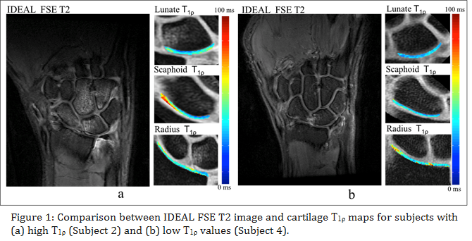
The mean scan/re-scan T1ρ CVs were 1.48%, 3.60% and 5.62% for the lunate, scaphoid and radial cartilage. The overall CV was 3.59%. In Tab. 1, the mean cartilage T1ρ values and MRI scores are reported. Strong linear correlations were found between the overall T1ρ values and both BMEL (R2 = 0.87, p<0.01) and erosion scores (R2 = 0.95, p<0.01). Fig. 1 shows FSE T2 IDEAL sequence and the corresponding T1ρ map of representative RA cases with high T1ρ (43.2 ms) and low T1ρ (36.9 ms).
Conclusion
We demonstrated excellent in vivo reproducibility of MRI T1ρ quantification in wrist cartilage. Despite the small sample size, the results obtained demonstrate the feasibility of using T1ρ to study the progression of cartilage damage and response to therapy in the wrist of RA patients.
References
[ = 1 * Arabic 1] Li et al, Osteoarthr Cartilage 2007 [2] Li et al, JMRI. 2012 [3] Crowley AR et al, JMRI. 2011
Disclosure:
V. Pedoia,
None;
F. Su,
None;
A. J. Burghardt,
None;
J. Graft,
None;
J. B. Imboden,
None;
M. Nakamura,
None;
U. Heilmeier,
None;
T. M. Link,
None;
X. Li,
None.
« Back to 2014 ACR/ARHP Annual Meeting
ACR Meeting Abstracts - https://acrabstracts.org/abstract/feasibility-and-clinical-implication-of-radiocarpal-cartilage-t1%cf%81-mr-imaging-in-rheumatoid-arthritis/


