ACR Meeting Abstracts

ACR Meeting Abstracts

  • Meetings
    • ACR Convergence 2024
    • ACR Convergence 2023
    • 2023 ACR/ARP PRSYM
    • ACR Convergence 2022
    • ACR Convergence 2021
    • ACR Convergence 2020
    • 2020 ACR/ARP PRSYM
    • 2019 ACR/ARP Annual Meeting
    • 2018-2009 Meetings
    • Download Abstracts
  • Keyword Index
  • Advanced Search
  • Your Favorites
    • Favorites
    • Login
    • View and print all favorites
    • Clear all your favorites
  • ACR Meetings

Abstract Number: 241

Exploratory Factor Analysis Of a Parsimonious Medial Cartilage Damage Index Reveals One Factor Associated With Radiographic Severity and Symptoms: Data From The Osteoarthritis Initiative

Grace H. Lo1, Lori Lyn Price2,3, Ming Zhang4, Jeffrey B. Driban5, Daniel Harper4 and Timothy E. McAlindon6, 1VA HSR&D Center for Innovations in Quality, Effectiveness and Safety; Medical Care Line and Research Care Line; Department of Medicine, Michael E. DeBakey VA Medical Center, Baylor College of Medicine, Houston, TX, 2Institute for Clinical Research and Health Policy Studies, Tufts Medical Center, Boston, MA, 3Biostatistics Research Center, Institute for Clinical Research and Health Policy Studies, Tufts Medical Center, Boston, MA, 4Rheumotology, Tufts Medical Center, Boston, MA, 5Rheumatology, Tufts Medical Center, Boston, MA, 6Division of Rheumatology, Tufts Medical Center, Boston, MA

Meeting: 2013 ACR/ARHP Annual Meeting

Keywords: cartilage and osteoarthritis, MRI

  • Tweet
  • Click to email a link to a friend (Opens in new window) Email
  • Click to print (Opens in new window) Print
Session Information

Title: Osteoarthritis - Clinical Aspects I: Risk Factors for and Sequelae of Osteoarthritis.

Session Type: Abstract Submissions (ACR)

Background/Purpose:

The cartilage damage index (CDI) is a parsimonious method of measuring articular cartilage approximating regional cartilage volume based on 18 locations within the knee, 9 in the medial tibia and femur (figure).  Understanding locations that are simultaneously pathologic may better clarify the pathophysiology of knee osteoarthritis (OA).  Therefore, we conducted an exploratory factor analysis of the CDI. 

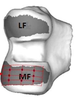
Figure. Spatial diagram of 9 of the femoral CDI points. Each red dot in the grid represents a separate location where CDI of articular cartilage is measured.

Methods:

We included a convenience sample of 200 participants of the progression cohort of the Osteoarthritis Initiative (OAI) with 3T knee MRIs, PA radiographs, and WOMAC pain scores (0 – 20) at the OAI 24- month visit.  Long limb films obtained at the OAI 12-, 24-, or 36- month visits were used to measure Hip-Knee-Ankle (HKA) angle using a semi-automated program (negative = varus).  One assessor performed CDI measurements on MRI sagittal DESS sequences.  Medial joint space narrowing (JSN) (0-3) and Kellgren-Lawrence (KL) score (0-4) were centrally assessed.  An exploratory factor analysis was conducted on the 18 CDI points.  To assess the characteristics of factors resulting from that analysis, we performed Spearman’s correlations with the full CDI measure, the separate factors, medial JSN (construct we are trying to capture), KL score (OA severity), HKA (risk factor for medial OA), and WOMAC score (OA symptoms). 

Results:

Mean age was 62.5 (9.5) years, mean BMI was 30.0 (4.5) kg/m2, 47% were female.  3 factors resulting from the factor analysis were: (1) anterior and weight bearing portions of the femur and tibia (AntWB) (2) posterior tibia (PostTib) and (3) posterior femur (PostFem), explaining 75%, 11%, and 9% of the data variation. 


CDI

AntWB CDI

PostTib CDI

PostFem CDI

Medial JSN

KL Score

HKA

WOMAC

CDI

1

0.97832

0.67338

0.68628

-0.50802

-0.28166

0.28489

-0.14135


<.0001

<.0001

<.0001

<.0001

<.0001

<.0001

0.0459

AntWB CDI


1

0.61494

0.56546

-0.57167

-0.35091

0.32417

-0.1724


<.0001

<.0001

<.0001

<.0001

<.0001

0.0146

PostTib CDI


1

0.45342

-0.1569

-0.10798

-0.04208

-0.04712


<.0001

0.0269

0.129

0.5581

0.5076

PostFem CDI


1

-0.1293

0.12819

0.11757

0.02053


0.0687

0.0712

0.1008

0.7729

Medial JSN


1

0.56874

-0.52189

0.09234


<.0001

<.0001

0.1946

KL Score


1

-0.19741

0.29629


0.0057

<.0001

HKA


1

0.02272


0.7519

WOMAC


1


Table. Spearman’s correlations of CDI, anterior weight-bearing (Ant WB) CDI, posterior tibia (PostTib) CDI, posterior femur (PostFem) CDI, Medial JSN, KL Score, HKA, and WOMAC scores.

Conclusion:  

These findings suggest 3 patterns of cartilage damage occur in the medial tibia and femur.  While PostTib and PostFem CDI are correlated with the full CDI, they are at best weakly correlated with medial JSN and not correlated with KL score, static alignment or pain.  The AntWB CDI pattern contributes most of the variation in the 18 points and shows construct validity, correlating with medial JSN, KL score, static alignment, and pain, better than the full CDI measurement.  Future studies confirming these findings are warranted.


Disclosure:

G. H. Lo,
None;

L. L. Price,
None;

M. Zhang,
None;

J. B. Driban,
None;

D. Harper,
None;

T. E. McAlindon,
None.

  • Tweet
  • Click to email a link to a friend (Opens in new window) Email
  • Click to print (Opens in new window) Print

« Back to 2013 ACR/ARHP Annual Meeting

ACR Meeting Abstracts - https://acrabstracts.org/abstract/exploratory-factor-analysis-of-a-parsimonious-medial-cartilage-damage-index-reveals-one-factor-associated-with-radiographic-severity-and-symptoms-data-from-the-osteoarthritis-initiative/

Advanced Search

Your Favorites

You can save and print a list of your favorite abstracts during your browser session by clicking the “Favorite” button at the bottom of any abstract. View your favorites »

All abstracts accepted to ACR Convergence are under media embargo once the ACR has notified presenters of their abstract’s acceptance. They may be presented at other meetings or published as manuscripts after this time but should not be discussed in non-scholarly venues or outlets. The following embargo policies are strictly enforced by the ACR.

Accepted abstracts are made available to the public online in advance of the meeting and are published in a special online supplement of our scientific journal, Arthritis & Rheumatology. Information contained in those abstracts may not be released until the abstracts appear online. In an exception to the media embargo, academic institutions, private organizations, and companies with products whose value may be influenced by information contained in an abstract may issue a press release to coincide with the availability of an ACR abstract on the ACR website. However, the ACR continues to require that information that goes beyond that contained in the abstract (e.g., discussion of the abstract done as part of editorial news coverage) is under media embargo until 10:00 AM ET on November 14, 2024. Journalists with access to embargoed information cannot release articles or editorial news coverage before this time. Editorial news coverage is considered original articles/videos developed by employed journalists to report facts, commentary, and subject matter expert quotes in a narrative form using a variety of sources (e.g., research, announcements, press releases, events, etc.).

Violation of this policy may result in the abstract being withdrawn from the meeting and other measures deemed appropriate. Authors are responsible for notifying colleagues, institutions, communications firms, and all other stakeholders related to the development or promotion of the abstract about this policy. If you have questions about the ACR abstract embargo policy, please contact ACR abstracts staff at [email protected].

Wiley

  • Online Journal
  • Privacy Policy
  • Permissions Policies
  • Cookie Preferences

© Copyright 2025 American College of Rheumatology