Session Information
Session Type: ACR Poster Session A
Session Time: 9:00AM-11:00AM
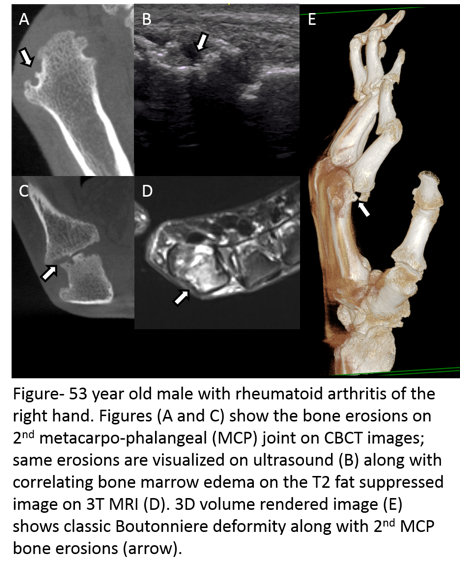
Background/Purpose: This preliminary study was designed to determine the agreement and reproducibility of a novel cone beam CT (CBCT) extremity scanner, MRI and ultrasound (US) for detection of erosions in rheumatoid arthritis (RA). For this purpose, we compared the bone erosion scores for CBCT, MRI, and US in RA patients along with test-retest reproducibility of the CBCT data.
Methods: Ten patients (5 males, 5 females; mean age 58 years, age range 34 – 81 years) with a clinically confirmed diagnosis of RA were recruited for this institutional review board-approved study. All patients underwent an MRI, CBCT and US scan of the wrist and hand followed by a second CBCT scan one week later. The MRI was done using a 3T clinical MR scanner with dedicated hand coil; US using a state-of-the-art machine with an 18 Mhz hockey stick probe; while the CBCT was done using a prototype CBCT extremity scanner. US scans were performed and read by a rheumatologist with 5 years of experience in musculoskeletal ultrasound, scoring for the presence of absence of erosion in each joint. A radiologist with 7 years of musculoskeletal imaging evaluated MRI and CBCT images for erosions using RAMRIS like scoring between 1 to 10 (1 is 0-10% erosion, 2 is 11-20% erosion etc). The 2nd to 5th metacarpophalangeal joints, carpal bones and distal radius and ulna were assessed. Repeatability and agreement between methods were evaluated with Intraclass Correlation Coefficients (ICC) and Bland-Altman plots.
Results: Bland-Altman analysis showed agreement of 0.02±3.5, 3.5 to -3.5 (bias ± repeatability coefficient, 95% limits of agreement interval) with ICC score of 0.77 showing good correlation between MRI, US, and CBCT bone erosion scores. Correlation was higher for bone erosion scores of MCPs than for wrist joints. Test-retest reproducibility for the CBCT scans showed an excellent ICC score of 0.95.
Conclusion: Good correlation was seen for bone erosions as detected by CBCT, MRI and US, while the prototype extremity CBCT images showed high test-retest reproducibility. This study provides a good first approximation for evaluation of bone erosions across the three modalities.
 Figure 1. Images of erosions on CBCT, MRI and US in an RA patient
To cite this abstract in AMA style:
Albayda J, Thawait G, Zbijewski W, Martin A, Demehri S, Fritz J, Yorkston J, Siewerdsen J, Bingham III CO. Evaluation of Bone Erosions in Rheumatoid Arthritis Patients Using Cone Beam Computed Tomography, Magnetic Resonance Imaging, and Ultrasound [abstract]. Arthritis Rheumatol. 2017; 69 (suppl 10). https://acrabstracts.org/abstract/evaluation-of-bone-erosions-in-rheumatoid-arthritis-patients-using-cone-beam-computed-tomography-magnetic-resonance-imaging-and-ultrasound/. Accessed .« Back to 2017 ACR/ARHP Annual Meeting
ACR Meeting Abstracts - https://acrabstracts.org/abstract/evaluation-of-bone-erosions-in-rheumatoid-arthritis-patients-using-cone-beam-computed-tomography-magnetic-resonance-imaging-and-ultrasound/
