Session Information
Date: Tuesday, October 23, 2018
Title: Osteoporosis and Metabolic Bone Disease – Basic and Clinical Science Poster
Session Type: ACR Poster Session C
Session Time: 9:00AM-11:00AM
Background/Purpose: Dual-energy X-ray absorptiometry (DXA) is recognized as the gold standard for measuring bone mineral density (BMD) with acceptable errors, good precision and reproducibility. However, the training of operators in different centers and countries is not standardized and the lack of knowledge can lead to errors both in the acquisition of information, as well as in its analysis and subsequent interpretation. The purpose is to determine the most common errors in the performance of bone densitometry from different imaging centers in Ecuador.
Methods: Cross-sectional descriptive study. We collected DXA scans from different imaging centers in Ecuador. Demographic information from patients included age, sex, height, weight, body mass index (BMI) and main diagnosis. Data from the DXA scan included city of origin, type of specialist that requested it and densitometry diagnosis. The DXA images provided were analyzed double blind by experts in the field from Argentina, according to patient position, presence of artifacts and region of interest correctly placed we.
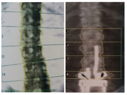
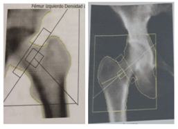
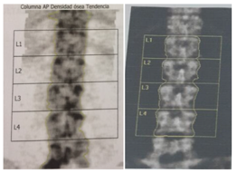
Results: From a total of 180 patients with a mean age of 63.5 years, 93.6% were women. 78% of the DXA scans came from private imaging centers and 22% from public centers, 95% of all came from the city of Guayaquil. The machines used were Hologic 51.2% and Lunar 48.8%. The densitometric diagnosis was 16.3% normal, 46.1% osteoporosis and 37.6% osteopenia. 112 left hip and 49 right hip scans were analyzed from which 31.1% and 22.4% had errors in patient positioning, respectively, mainly internal or external rotation. 140 lumbar scans were analyzed from which 21.4% had patient positioning errors (not centered or not straight). Also in 38.5% the vertebral area did not correspond to L1-L4. 3.5% had artefacts such as a metal bar or implant. The region of interest was misplaced in 24.1% of the lumbar scans and 19.9% of the femur.
Conclusion: Much of the responsibility of a DXA falls on the operator as he has to review the patient’s health history, enter demographic data, perform the acquisition of the image with a correct positioning and analyze it. When studies are performed incorrectly, it can lead to important errors in diagnosis and therapy.
Figure 1: Spine Incorrect alignment and prosthetics
Figure 2. Femur Incorrect vs. correct alignment
Figure 3. Spine Incorrect vs. correct alignment.
To cite this abstract in AMA style:
Maldonado G, Intriago MJ, Larroude M, Aguilar G, Moreno M, Gonzalez J, Vargas S, Vera C, Guerrero R, Ríos K, Rios C. Errors and Discrepancies in DXA Scans in Imaging Centers in Ecuador [abstract]. Arthritis Rheumatol. 2018; 70 (suppl 9). https://acrabstracts.org/abstract/errors-and-discrepancies-in-dxa-scans-in-imaging-centers-in-ecuador/. Accessed .« Back to 2018 ACR/ARHP Annual Meeting
ACR Meeting Abstracts - https://acrabstracts.org/abstract/errors-and-discrepancies-in-dxa-scans-in-imaging-centers-in-ecuador/


