Session Information
Date: Sunday, November 13, 2016
Title: Imaging of Rheumatic Diseases I: Advanced Imaging in RA and Spondyloarthritides
Session Type: ACR Concurrent Abstract Session
Session Time: 2:30PM-4:00PM
Background/Purpose: Rheumatoid arthritis (RA) can be differentiated according to rheumatoid factor (RF) and citrullinated peptide antibodies (ACPA). It is unclear whether seropositive RA (spRA) and seronegative RA (snRA) are different forms of the same disease or separate entities. Differing manifestation patterns between spRA and snRA on hand radiographs were first described by Burns/Calin in 1983[1]. To our knowledge, this observation has not been pursued further.
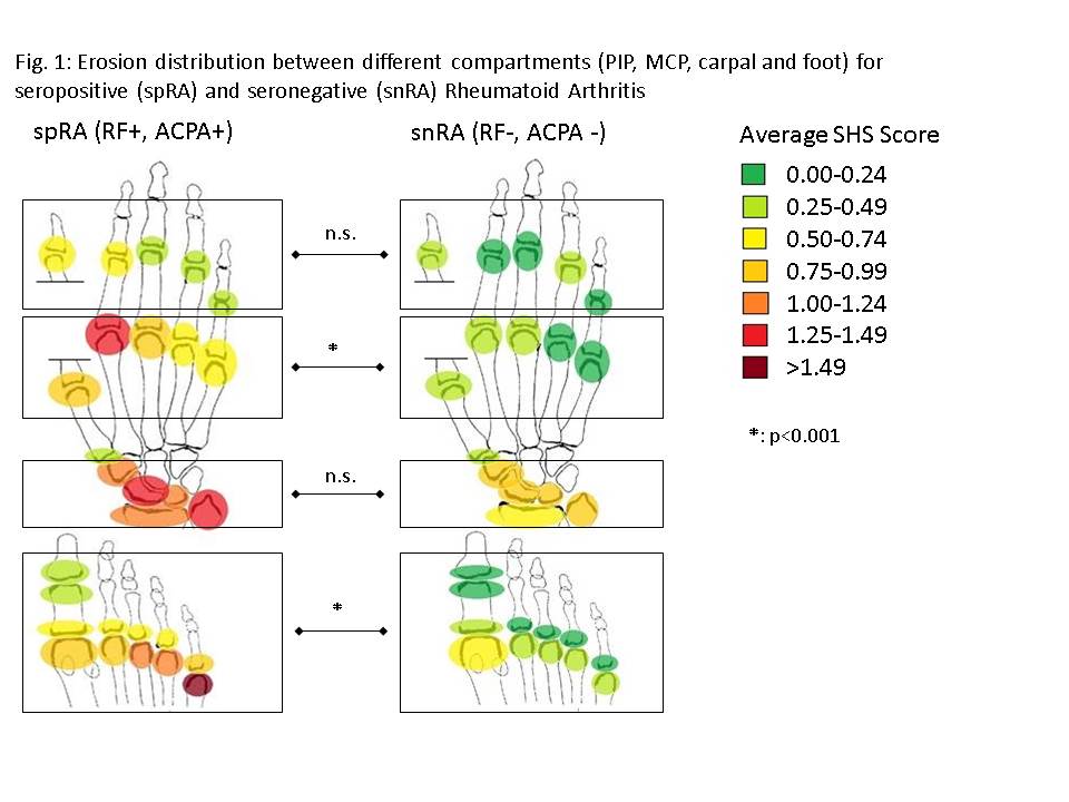
Methods: Hand and foot radiographs of 54 patients with RF negative, ACPA negative, erosive RA (snRA) were evaluated according to the Sharp-van-der-Heijde score (SHS) and compared with radiographs of 55 age-matched, RF-positive, ACPA-positive, erosive RA (spRA) patients. The average SHS for both cohorts was determined for the entire set of radiographs, for the carpal, MCP, PIP and foot compartments and for each single joint separately. We also evaluated the relative erosion score for each compartment (compartment SHS/total SHS). The results were transferred to a ‘heat map’ for visual representation. All data are given as mean± SD.
Results: Total SHS was significantly higher in spRA than in snRA (SHS 47.4± 53.5 vs. 20.6± 24.7, p=0.0012). The difference in SHS was significant in the MCP (8.5± 11.3 vs. 2.6± 4.7, p<0.001) and foot (20.9± 26.3 vs. 6.1± 14.8, p<0.001) but not in the PIP (4.9± 9.7 vs. 2.2± 5.4) or carpal (12.2± 14.4 vs. 8.8± 12.5, p=0.192) compartments (Fig.1). The greatest single-joint difference was found in MCP 2 (1.26± 1.60 vs. 0.27± 0.48) and proximal MTP 5 (2.11± 1.48 vs. 0.44± 0.89, p<0.0001 respectively). Significant differences in relative erosion score were found in the carpal (28± 23% vs. 42± 33%, p=0.017) and foot (44± 26% vs. 27± 34%, p=0.004) compartments.
Conclusion: Sharp-van-der-Heijde score (SHS) is higher in seropositive RA (spRA) than in seronegative RA (snRA). The difference in SHS is significant for the MCP and foot, but not for the carpal or PIP compartments. The single joint difference is greatest in MCP 2 and proximal MTP 5. Considering the relative erosion score (compartment SHS/total SHS), spRA shows a predilection for the foot and snRA for the carpal compartment. The different erosion patterns suggest that pathophysiological processes differ between the forms. 
[1] Burns T, Calin A. The hand radiograph as a diagnostic discriminant between seropositive and seronegative ‘rheumatoid arthritis’: a controlled study. Annals of the Rheumatic Diseases, 1983, 42, 605-612
To cite this abstract in AMA style:
Gadeholt O, Hausotter K, Eberle H, Tony HP, Schmalzing M, Klink T. Erosion Patterns in Seropositive and Seronegative Rheumatoid Arthritis: A Joint-By-Joint Approach [abstract]. Arthritis Rheumatol. 2016; 68 (suppl 10). https://acrabstracts.org/abstract/erosion-patterns-in-seropositive-and-seronegative-rheumatoid-arthritis-a-joint-by-joint-approach/. Accessed .« Back to 2016 ACR/ARHP Annual Meeting
ACR Meeting Abstracts - https://acrabstracts.org/abstract/erosion-patterns-in-seropositive-and-seronegative-rheumatoid-arthritis-a-joint-by-joint-approach/
