Session Information
Date: Thursday, March 19, 2026
Title: Abstracts: Imaging
Session Time: 5:35PM-5:40PM
Background/Purpose: JDM and childhood-onset SLE (cSLE) are systemic autoimmune disorders with pediatric onset. While interferon-driven inflammation is a key signature that affects multiple organs across both conditions, central nervous system involvement is more clinically prevalent in cSLE, although still vastly understudied in JDM. Therefore, in this pilot study we aim to (i) explore structural brain abnormalities in a combined sample of adolescents diagnosed with JDM or cSLE and no clinically documented neuropsychiatric symptoms, and (ii) to evaluate if these associate with disease features such as activity, duration, and glucocorticoid treatment, and self-reported measures of executive function.
Methods: Structural magnetic resonance imaging (MRI) was cross-sectionally acquired at 3T in 10 adolescents with JDM, 15 with cSLE and 25 age/sex comparable healthy controls (HC) aged 10-18 years. For each participant, total white matter (WM) hyperintensities, cortical and subcortical grey matter (GM), WM, and cerebrospinal fluid volumes were automatically segmented with AssemblyNet pipeline (Fig. 1). Global brain age was estimated with BrainStructureAges pipeline and the difference (gap) between participant’s global brain age and chronological age was calculated (Fig. 1). The Behavioral, Emotional, and Cognitive Regulation Indices, and the Global Executive Composite from the Behavior Rating Inventory of Executive Function questionnaire evaluated self-reported executive function. Group differences in clinical and brain metrics were calculated with analyses of variance/covariance, and associations between brain and cognitive metrics were tested with Pearson correlations.
Results: In our combined disease sample, JDM participants were diagnosed at younger ages and had higher cumulative glucocorticoid doses, likely due to longer disease duration when compared to youth with cSLE, although most patients (84%) were not on current glucocorticoid treatment (Table 1). Brain volumetric group/subgroup analyses revealed higher T2-weighted WM hyperintensities volume and lower subcortical GM volume in patients compared to HC, although not specific for any disease subgroup (Fig 2A, B), while greater positive brain age gaps in patients versus HC were significantly driven by JDM patients (Fig. 2C). Compared to HC, adolescents with JDM and cSLE did not show differences on any self-reported executive function scores (Table 1). Greater brain age gaps in all patients were positively correlated with worse (elevated) emotional self-regulation (Fig. 2D), and these associations strengthened when adjusting for the effects of disease duration and cumulative glucocorticoid doses.
Conclusion: Adolescents diagnosed with inflammatory autoimmune disorders such as JDM and cSLE exhibited brain structural abnormalities compared to healthy peers, and higher brain age gaps correlating to worse self-reported executive function. Accelerated brain aging, possibly as a consequence from mild global/local brain atrophy, affects aspects of executive function, may be aggravated by the cumulative effects of chronic disease and/or glucocorticoids, and requires future longitudinal research in larger samples.
Figure 1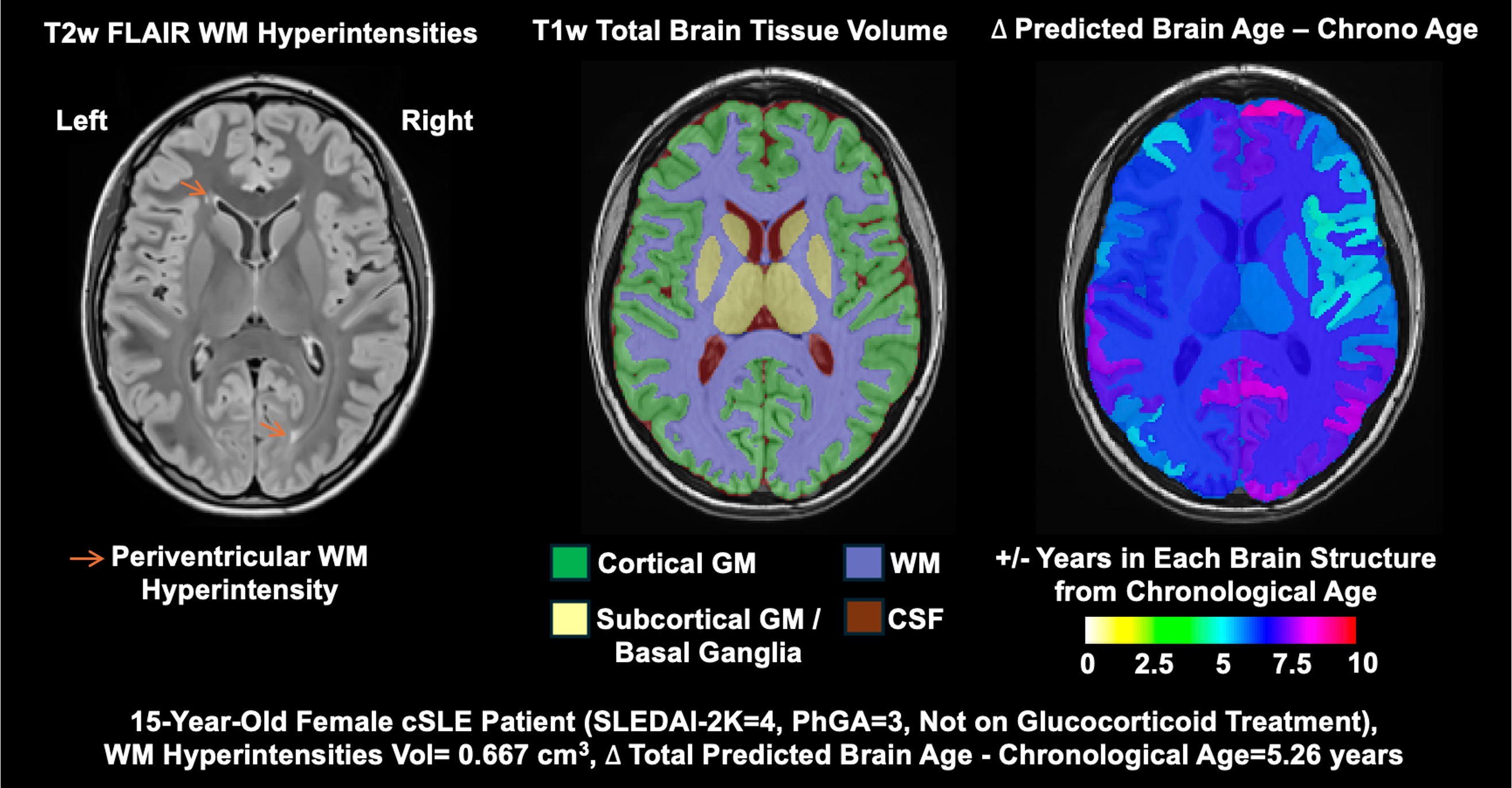 Axial MRI views (neurological convention) of a representative cSLE patient example showing white matter (WM) hyperintensities in anterior and posterior periventricular regions (left, T2-weighted Fluid-Attenuated Inversion Recovery, FLAIR MRI); total WM, cortical and subcortical grey matter (GM), and cerebrospinal fluid (CSF) tissue volume segmentations (middle, T1-weighted MRI); and predicted brain ages across different local structures minus chronological age (right, T1-weighted MRI), these regional brain ages are utilized to calculate the global brain age difference or gap. For this patient, a global brain age gap of 5.26 years is indicative of accelerated brain aging.
Table 1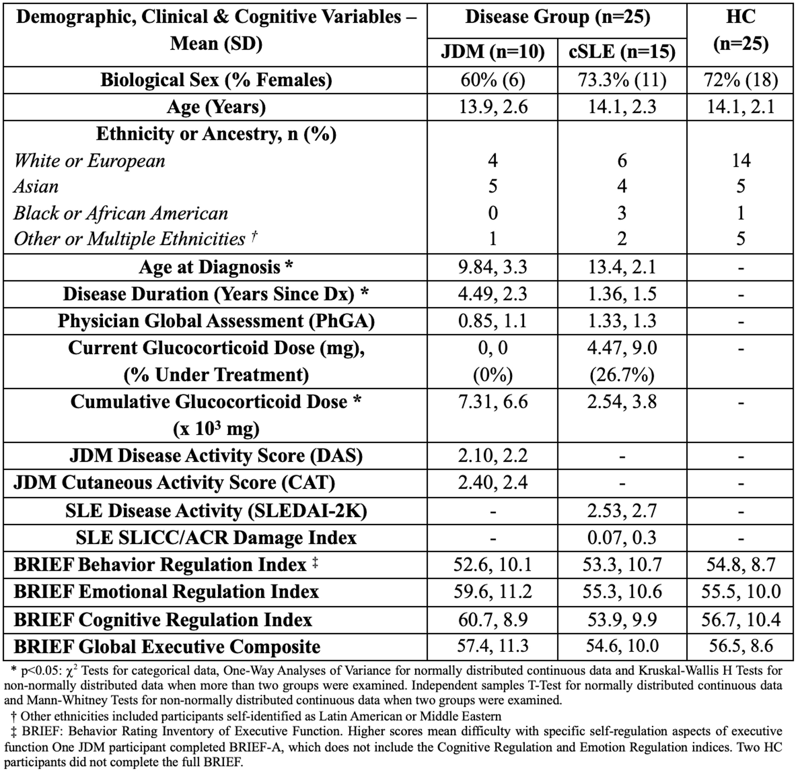 Demographics and Clinical Characteristics, and Self-Reported Executive Function T-Scores of JDM, cSLE, and HC groups.
Figure 2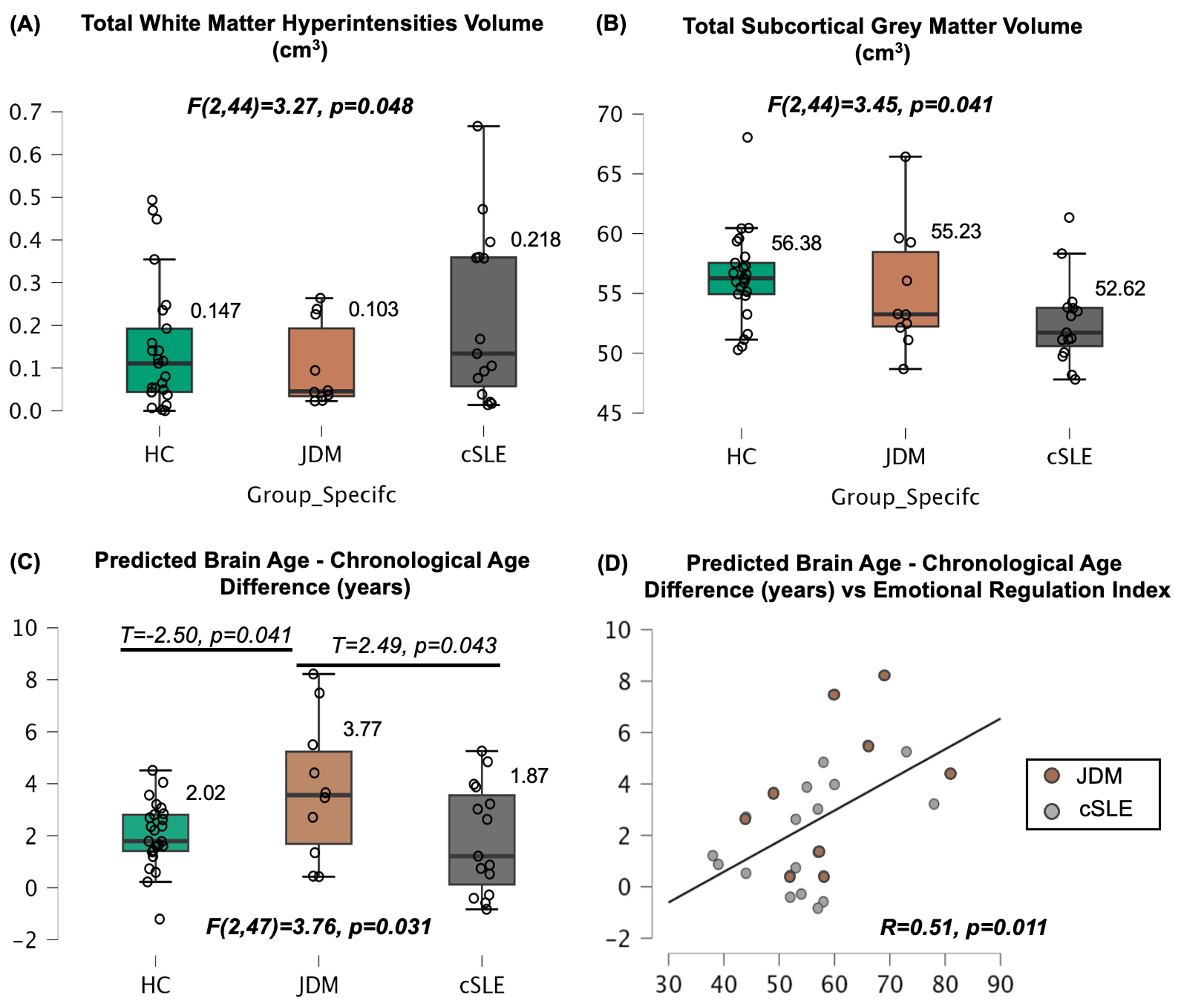 Patients exhibited higher white matter hyperintensities (A), lower subcortical GM (B), and higher brain age gaps/differences (C) compared to HC. Greater brain age gaps were significantly driven by patients diagnosed with JDM. In patients, greater brain age gaps were positively associated with worse emotional regulation indices (D).
To cite this abstract in AMA style:
Valdes Cabrera D, Tohan E, Jeyanathan A, Jaffan J, Mossad S, Wagner B, Feldman B, Knight A. Effects of Childhood-Onset Interferon-Driven Autoimmune Disorders on Brain Volume and Executive Function: A Pilot Analysis in JDM and Childhood-Onset SLE [abstract]. Arthritis Rheumatol. 2026; 78 (suppl 3). https://acrabstracts.org/abstract/effects-of-childhood-onset-interferon-driven-autoimmune-disorders-on-brain-volume-and-executive-function-a-pilot-analysis-in-jdm-and-childhood-onset-sle/. Accessed .« Back to 2026 Pediatric Rheumatology Symposium
ACR Meeting Abstracts - https://acrabstracts.org/abstract/effects-of-childhood-onset-interferon-driven-autoimmune-disorders-on-brain-volume-and-executive-function-a-pilot-analysis-in-jdm-and-childhood-onset-sle/