Session Information
Date: Tuesday, November 12, 2019
Title: Osteoporosis & Metabolic Bone Disease – Basic & Clinical Science Poster
Session Type: Poster Session (Tuesday)
Session Time: 9:00AM-11:00AM
Background/Purpose: A novel, non-ionizing approach – Radiofrequency Echographic Multi Spectrometry (REMS) was recently introduced for diagnosis of osteoporosis on both sites-lumbar spine and femoral neck. In the previous studies, bone mineral density (BMD) assessed by REMS technique highly correlated with BMD obtained by dual X-ray absorptiometry (DXA). However, there is no previous study, which investigated the diagnostic role of the dual femur BMD measurements based on REMS technology. The aim of this study is to compare the REMS-based BMD values of both femora-left and right in postmenopausal women and to identify the number of the women, in which the inclusion of bilateral hip in BMD measurement changed the diagnosis to more severe.
Methods: Dual femur was assessed in 72 postmenopausal women with mean age 61 years ± 13 standard deviations (SD), (range 40-66 years). Data was analyzed with SPSS version 21.0. Patients were classified as osteopenic if -2.5 SD < T-score< -1.0 SD, as osteoporotic if T-score ≤ -2.5 SD and healthy if T-score ≥-1.0. Linear regression analysis was used to compare the REMS-based BMD values of both femora.
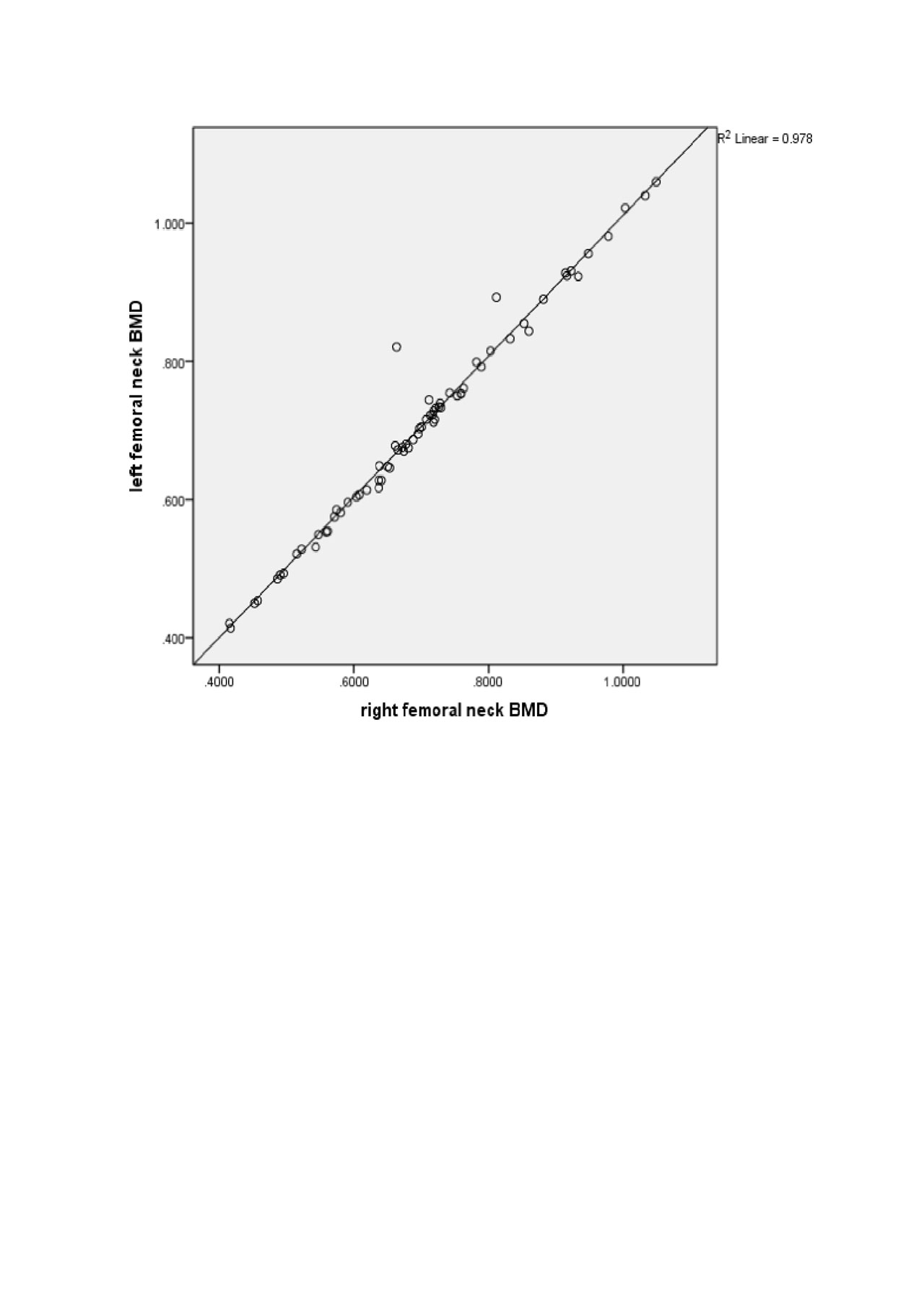
Results: Based on the unilateral (left) hip BMD measurements, 22 women were diagnosed with normal BMD, 35- with osteopenia and 15 – with osteoporosis. The mean left femoral neck BMD was 0.879 g/cm2 ± 0.100 g/cm2 in the women with normal BMD, 0.685 g/cm2 ± 0.056 g/cm2 in the women with osteopenia and 0.507 g/cm2 ± 0.054 g/cm2 in the women with osteoporosis. The mean right femoral neck BMD was 0.873 g/cm2 ± 0.095 g/cm2 in the women with normal BMD, 0.677 g/cm2 ± 0.046 g/cm2 in the women with osteopenia and 0.507 g/cm2 ± 0.054 g/cm2 in the women with osteoporosis. A strong correlation between left and right femoral neck BMD values was observed (r2=0.978, p=0.000), (figure 1). 63.6% (14/22) of the women with normal BMD had a T-score discordance equal to 0.1 and 4.6% (1/22) – equal to 0.2. 31.8% (7/22) of the women with normal BMD were attributed to a more severe diagnosis – osteopenia after assessing the dual femur. 48.6% (17/35) of the women with osteopenia had a T-score discordance equal to 0.1, 8.6% (3/35) – equal to 0.2 and 2.9% (1/35) – equal to 0.5. 2.9% (1/35) of the women with osteopenia were attributed to a more severe diagnosis – osteoporosis after assessing the dual femur. 33.3% (5/15) of the women with osteoporosis showed a T-score discordance equal to 0.1.
Conclusion: In women with femoral neck BMD values, which correspond to borderline T-scores, dual femur BMD measurements could be crucial for obtaining a more accurate diagnosis and decision about the treatment.
To cite this abstract in AMA style:
Kirilova E, Kirilov N, Nikolov M, Nikolov N, Popov I, Vladeva S. Dual Femur Bone Density Measurements with Novel Sonographic Approach – Radiofrequency Echographic Multi Spectrometry [abstract]. Arthritis Rheumatol. 2019; 71 (suppl 10). https://acrabstracts.org/abstract/dual-femur-bone-density-measurements-with-novel-sonographic-approach-radiofrequency-echographic-multi-spectrometry/. Accessed .« Back to 2019 ACR/ARP Annual Meeting
ACR Meeting Abstracts - https://acrabstracts.org/abstract/dual-femur-bone-density-measurements-with-novel-sonographic-approach-radiofrequency-echographic-multi-spectrometry/

