Session Information
Session Type: Abstract Submissions (ACR)
Background/Purpose: To better characterize the soft tissue details of systemic sclerosis–related (SSc) calcinosis of the hands using dual-energy computer tomography (DECT). DECT is an imaging modality used similarly to study monosodium urate (MSU) deposition in gout (1). Methods: Fourteen patients with symptomatic hand SSc-calcinosis had DECT imaging and their clinical characteristics reviewed.
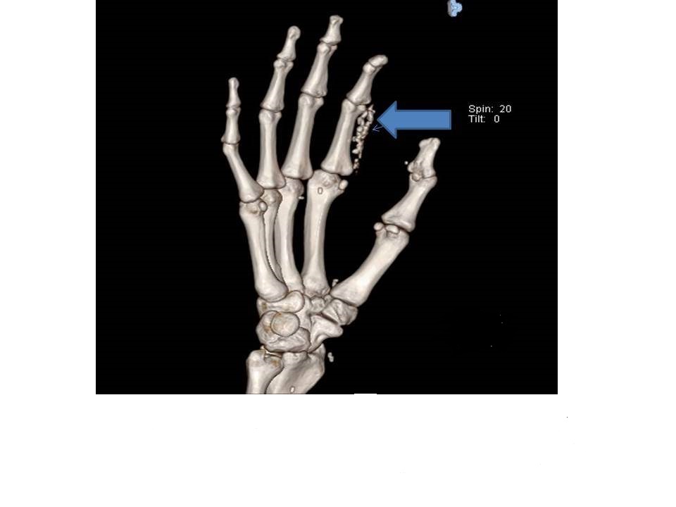
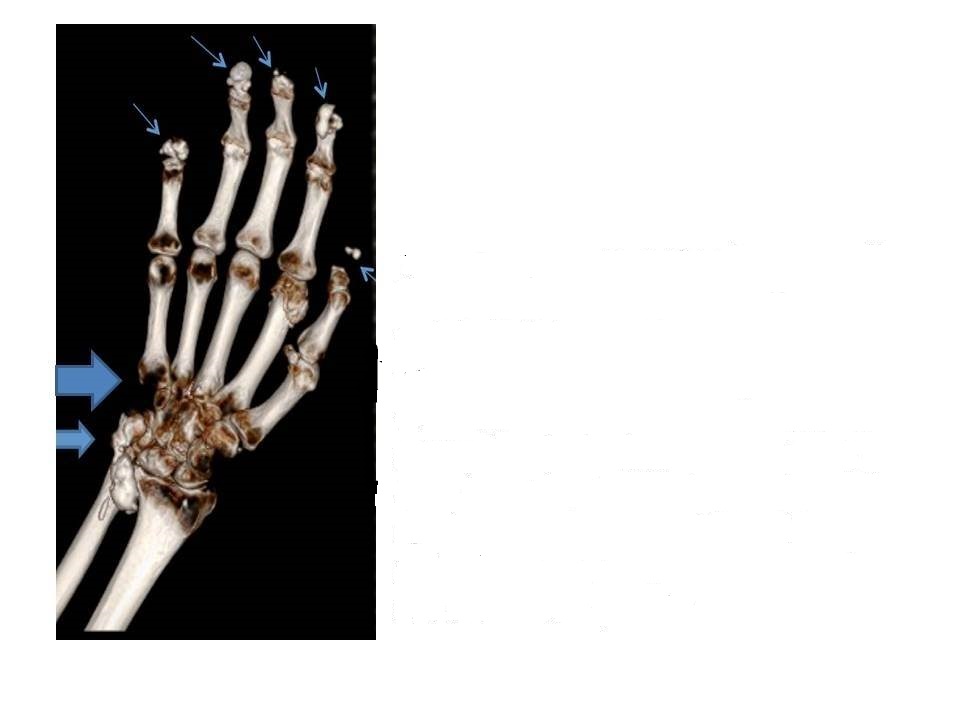
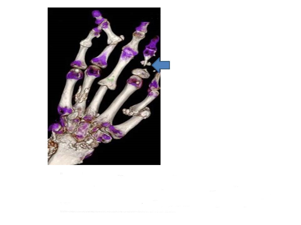
Results: Eight of 14 patients had diffuse SSc and 6 also had rheumatoid arthritis (RA). Herein are 3 DECT images: Image 1: a caucasian male with lifelong calcinosis due to diffuse scleroderma, whose painful deposits drain intermittently. Image 2: a caucasian female with limited SSc and RA for 15 years, whose calcinosis was found by imaging. Image 3: an African-American female with diffuse scleroderma and RA for 10 years. Her calcinosis caused painful wrist swelling, but despite surgery, continues to accumulate.
On DECT imaging, calcinosis was most commonly found in the subcutaneous (SQ) fat pads of the fingertips and along tendon sheaths and muscle groups. Acro-osteolysis was present in most (93%) patients, 7 (50%) with calcinosis nearby. No MSU was identified. Our cohort also had pulmonary fibrosis (79%), ischemic digital ulcers (71%) and bowel (43%) complications. Conclusion: SSc-calcinosis affects diffuse and limited SSc. We found DECT imaging better defined the soft tissue details and was useful in the evaluation of SSc-calcinosis. References:
1. Choi K: Ann Rheum Dis 2009; 68 (10):1609.

Image 1: 3D image shows acro-osteolysis of the 2nd and 3rd digits, extensive calcification in the SQ fat (volar) of the second digit (arrow), punctate deposits in the fat pad of thumb, lateral to the scaphoid bone, and between several MCP joints.
Image 2: Acro-osteolysis with associated calcinosis in the fat pads of all fingertips (small arrows); extensive calcification seen around the 1st and 2nd MCP and ulna (adjacent to the carpal tunnel) (medium arrow). The distal 5th phalanx is completely eroded (large arrow). The wrist deposits appear separate from the tendons.
Image 3: Soft tissue calcifications seen throughout the fingers and wrist with marked destruction at the radio-carpal joint. The scaphoid may be partially collapsed. There is severe penciling with nearly complete bone resorption (large arrow) of mid portion of 2nd phalanx.
Disclosure:
V. Hsu,
None;
M. Bramwit,
None;
N. Schlesinger,
Novartis Pharmaceutical Corporation,
2,
Takeda,
8,
Sobi,
9,
Astra Zeneca ,
9.
« Back to 2014 ACR/ARHP Annual Meeting
ACR Meeting Abstracts - https://acrabstracts.org/abstract/dual-energy-computed-tomography-for-the-evaluation-of-calcinosis-in-systemic-sclerosis/
