Session Information
Date: Tuesday, October 28, 2025
Title: (2524–2546) Vasculitis – Non-ANCA-Associated & Related Disorders Poster III
Session Type: Poster Session C
Session Time: 10:30AM-12:30PM
Background/Purpose: Several studies have demonstrated increased venous wall thickness (VWT) in patients with Behçet’s syndrome (BS) compared to healthy and diseased control groups. Ultrasonographic measurement of VWT has been proposed as a potential diagnostic tool to differentiate BS from other diseases. However, the diagnostic utility of this method remains controversial. An optimal approach to evaluate the diagnostic value of VWT measurement would be to perform Doppler ultrasonography in patients referred for screening with a suspicion of BS, and determining the sensitivity and specificity based on the measurements of those who are and who are not diagnosed with BS after a multidisciplinary diagnostic work-up.
Methods: All individuals who presented to our clinic for BS screening between May 2023 and December 2023 were included. Additionally, 30 patients with vascular BS and deep vein thrombosis (DVT) and 36 healthy individuals were also enrolled. Two radiologists, blinded to the clinical diagnoses, independently measured the wall thickness of the common femoral vein (CFV), great saphenous vein (GSV), and small saphenous vein (SSV). Results were presented as medians and interquartile ranges, and intergroup comparisons were conducted using the Kruskal–Wallis test.
Results: Among 84 screened individuals, BS was diagnosed in 45 patients, while 39 were found to have non-BS conditions. Lower extremity DVT was present in 5 of the BS patients and in 2 of the non-BS patients. The non-BS group were diagnosed with other conditions including spondyloarthritis, uveitis, inflammatory bowel disease, recurrent aphthous stomatitis, connective tissue diseases, systemic autoinflammatory disorders, and chronic thromboembolic pulmonary hypertension. Compared to other groups, healthy controls were younger, had lower body mass index (BMI), and were less frequently smokers. The highest median CFV thickness was observed in vascular BS patients [right: 0.76 mm (0.63–0.87), left: 0.71 mm (0.62–0.86)], followed by patients diagnosed with BS after screening [right: 0.59 mm (0.53–0.69), left: 0.60 mm (0.50–0.68)], non-BS patients diagnosed after screening [right: 0.58 mm (0.50–0.66), left: 0.57 mm (0.49–0.67)], and healthy controls [right: 0.49 mm (0.47–0.53), left: 0.48 mm (0.43–0.50)]. CFV thickness was significantly higher in vascular BS patients and significantly lower in healthy controls (p < 0.001 for all comparisons). However, there was no significant difference between BS and non-BS patients, and this pattern was consistent across other examined veins. This pattern remained unchanged after excluding the 7 patients with a history of lower extremity DVT. Inter-observer reliability between the radiologists was excellent (ICC for right CFV: 0.97; left CFV: 0.96).
Conclusion: Venous wall thickness was higher in both BS and non-BS patients compared to healthy individuals. However, VWT measurement did not aid in differentiating BS from other inflammatory diseases in patients referred for BS evaluation. The significantly increased VWT in vascular BS raises the question of whether this finding reflects vascular inflammation or vascular remodeling.
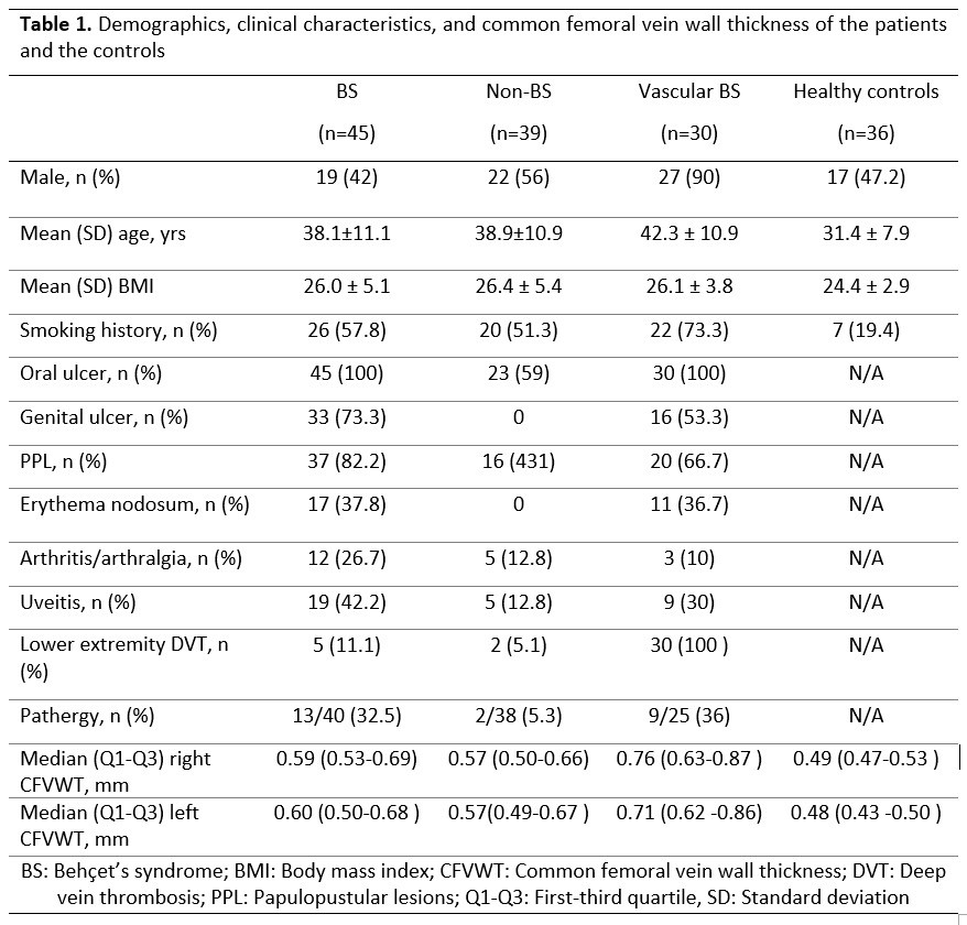 Table 1. Demographics, clinical characteristics, and common femoral vein wall thickness of the patients and the controls
.jpg) Figure 1. Right and left common femoral vein wall thickness across diagnostic groups
Figure 1. Right and left common femoral vein wall thickness across diagnostic groups
To cite this abstract in AMA style:
Alcin Bayraktar r, Kayadibi Y, Kahveci A, Esatoglu S, Adaletli I, Ozguler Y, Melikoglu M, Hatemi G. Diagnostic value of vein wall thickness measurement for patients with suspected Behçet syndrome [abstract]. Arthritis Rheumatol. 2025; 77 (suppl 9). https://acrabstracts.org/abstract/diagnostic-value-of-vein-wall-thickness-measurement-for-patients-with-suspected-behcet-syndrome/. Accessed .« Back to ACR Convergence 2025
ACR Meeting Abstracts - https://acrabstracts.org/abstract/diagnostic-value-of-vein-wall-thickness-measurement-for-patients-with-suspected-behcet-syndrome/