Session Information
Date: Tuesday, November 12, 2019
Title: Vasculitis – Non-ANCA-Associated & Related Disorders Poster III: Giant Cell Arteritis
Session Type: Poster Session (Tuesday)
Session Time: 9:00AM-11:00AM
Background/Purpose: In addition to aiding in diagnosis, histopathologic findings from temporal artery biopsy (TAB) specimens in giant cell arteritis (GCA) may be valuable for their associations with clinical features of the disease. The study objective was to compare specific histopathologic findings on TAB with biopsy interpretation, demographic, clinical, and imaging features at diagnosis.
Methods: Patients who had a diagnosis of GCA confirmed by expert review were selected from an international, multicenter observational cohort. Associations between demographic, clinical, radiographic, and histopathologic features were identified using bivariate testing and multivariate regression modeling.
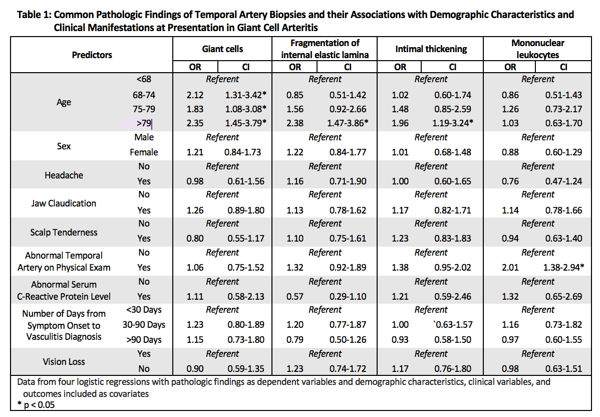
Results: Six hundred forty-seven patients with GCA who underwent TAB were included. Pathology interpretations included definite vasculitis (69%), normal temporal artery biopsy (13%), consistent with vasculitis but not definite (9%), or non-diagnostic (8%). For 467 biopsies, specific findings were detailed, including the presence of giant cells (63%), fragmentation of the internal elastic lamina (50%), intimal thickening (39%), predominantly mononuclear leukocytes (39%), vascular thrombosis (6%), granuloma (5%), and perivascular inflammation without vessel wall involvement (1%). In a model that included the common TAB findings ( >10% prevalence), a pathology interpretation of definite vasculitis was independently associated with giant cells (OR 123.4, CI 47.1-323.5), predominantly mononuclear leukocytes (OR 11.8, CI 6.0-23.0), and fragmentation of the internal elastic lamina (OR 4.5, CI 2.4-8.3). Out of 207 patients who had a temporal artery ultrasound, 166 patients had a halo sign, which was significantly associated with the presence of giant cells (88% vs. 76% without, p=0.04) and intimal thickening (90% vs. 78% without, p = 0.04). Out of 70 patients who underwent PET scanning, no pathologic findings on TAB were associated with PET abnormalities. Temporal artery abnormalities on exam were associated mononuclear leukocytes (OR 2.01, CI 1.38-2.94). Older age was associated with the presence of giant cells, fragmentation of the internal elastic lamina, and intimal thickening (see Table 1). No pathologic findings were associated with vision loss at presentation.
Conclusion: Pathologic findings of temporal artery biopsies in GCA are associated with diagnostic interpretations but not with clinical manifestations of vasculitis. Associations between pathologic findings and imaging abnormalities should be investigated further.
To cite this abstract in AMA style:
Putman M, Gribbons K, Craven A, Ponte C, Robson J, Suppiah R, Watts R, Luqmani R, Merkel P, Archer A, Grayson P. Clinicopathologic Associations in a Large International Cohort of Patients with Giant Cell Arteritis [abstract]. Arthritis Rheumatol. 2019; 71 (suppl 10). https://acrabstracts.org/abstract/clinicopathologic-associations-in-a-large-international-cohort-of-patients-with-giant-cell-arteritis/. Accessed .« Back to 2019 ACR/ARP Annual Meeting
ACR Meeting Abstracts - https://acrabstracts.org/abstract/clinicopathologic-associations-in-a-large-international-cohort-of-patients-with-giant-cell-arteritis/

