ACR Meeting Abstracts

ACR Meeting Abstracts

  • Meetings
    • ACR Convergence 2024
    • ACR Convergence 2023
    • 2023 ACR/ARP PRSYM
    • ACR Convergence 2022
    • ACR Convergence 2021
    • ACR Convergence 2020
    • 2020 ACR/ARP PRSYM
    • 2019 ACR/ARP Annual Meeting
    • 2018-2009 Meetings
    • Download Abstracts
  • Keyword Index
  • Advanced Search
  • Your Favorites
    • Favorites
    • Login
    • View and print all favorites
    • Clear all your favorites
  • ACR Meetings

Abstract Number: 217

Clinical Value Of 18f-Fluoro-Dexoxyglucose Positron Emission Tomography In Patients With Adult-Onset Still’s Disease

Hiroyuki Yamashita1, Kazuo Kubota2, Yuko Takahashi1, Hiroshi Kaneko1, Toshikazu Kano1 and Akio Mimori1, 1Division of Rheumatic Diseases, National Center for Global Health and Medicine, Tokyo, Japan, 2Department of Radiology, National Center for Global Health and Medicine, Tokyo, Japan

Meeting: 2013 ACR/ARHP Annual Meeting

Keywords: Adult-onset Still's disease

  • Tweet
  • Email
  • Print
Session Information

Title: Imaging of Rheumatic Diseases I: Imaging in Gout, Pediatric, Soft and Connective Tissue Diseases

Session Type: Abstract Submissions (ACR)

Abstract

Background/Purpose: The aim of this study was to assess the usefulness of 18F-fluoro-dexoxyglucose positron emission tomography/Computed tomography (18F-FDG PET/CT) for the diagnosis or the evaluation of Adult-onset Still’s disease (AOSD) visually.

Methods: 7 consecutive AOSD patients who had undergone PET at our department between 2007 and 2012 were included in the study. In addition, the literature review was performed about 6 previously reported AOSD patients who had undergone PET. We evaluate FDG uptake for characteristic findings in patients with AOSD.

Results: FDG accumulation was positive mainly in the bone marrow (70%), spleen (80%), lymph nodes (77.8%), and joints (50%). In addition, FDG uptake was positive in the pericardium, pleura, salivary glands, eyelids, muscle, and major blood vessels. 8 patients underwent FDG PET/CT for evaluating the efficacy of treatment. The follow-up PET showed diminished FDG accumulation in the bone marrow, spleen, lymph nodes , with SUVmax being significantly reduced from 4.03±0.95 to 2.20±0.75 (P=0.04), from 4.15±1.10 to 2.55±1.13(P=0.04), and from 5.47±5.19 to 2.10±1.91 (P=0.11), respectively. No significant correlation was found between max SUVs in each site and the laboratory data; the only significant correlation was between LDH and the spleen SUV.

Conclusion: Our study suggests that 18F-FDG PET may play a potential role in diagnosing or monitoring of disease activity marker in patients with AOSD. Additionally,we suspect that future PET/CT imaging will reveal that AOSD is a disease involving lesions with a little clinical description.

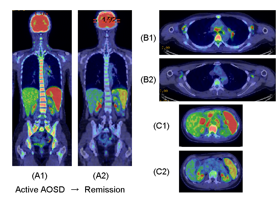
Fig.1FDG-PET/CT images at diagnosis (A1,B1,C1) and after steroid and Tocilizmab treatment (A2,B2,C2) in a 32-year-old woman (Patient 4) with AOSD.

Marked FDG accumulation was observed in the bone marrow, spleen and multiple lymph nodes at the time of diagnosis before treatment. After treatment, bone marrow FDG uptake improved from SUVmax��4.02 (A1) to 2.50 (A2) and spleen FDG uptake decreased from SUVmax��6.05(A1,C1) to 4.38(A2,C2). In addition, multiple lymph node FDG uptake observed in the axilla, mediastinum, hilar region of the lung, hilar region of the liver and para-aortic region declined or disappeared (B1/C1→B2/C2).


Disclosure:

H. Yamashita,
None;

K. Kubota,
None;

Y. Takahashi,
None;

H. Kaneko,
None;

T. Kano,
None;

A. Mimori,
None.

  • Tweet
  • Email
  • Print

« Back to 2013 ACR/ARHP Annual Meeting

ACR Meeting Abstracts - https://acrabstracts.org/abstract/clinical-value-of-18f-fluoro-dexoxyglucose-positron-emission-tomography-in-patients-with-adult-onset-stills-disease/

Advanced Search

Your Favorites

You can save and print a list of your favorite abstracts during your browser session by clicking the “Favorite” button at the bottom of any abstract. View your favorites »

All abstracts accepted to ACR Convergence are under media embargo once the ACR has notified presenters of their abstract’s acceptance. They may be presented at other meetings or published as manuscripts after this time but should not be discussed in non-scholarly venues or outlets. The following embargo policies are strictly enforced by the ACR.

Accepted abstracts are made available to the public online in advance of the meeting and are published in a special online supplement of our scientific journal, Arthritis & Rheumatology. Information contained in those abstracts may not be released until the abstracts appear online. In an exception to the media embargo, academic institutions, private organizations, and companies with products whose value may be influenced by information contained in an abstract may issue a press release to coincide with the availability of an ACR abstract on the ACR website. However, the ACR continues to require that information that goes beyond that contained in the abstract (e.g., discussion of the abstract done as part of editorial news coverage) is under media embargo until 10:00 AM ET on November 14, 2024. Journalists with access to embargoed information cannot release articles or editorial news coverage before this time. Editorial news coverage is considered original articles/videos developed by employed journalists to report facts, commentary, and subject matter expert quotes in a narrative form using a variety of sources (e.g., research, announcements, press releases, events, etc.).

Violation of this policy may result in the abstract being withdrawn from the meeting and other measures deemed appropriate. Authors are responsible for notifying colleagues, institutions, communications firms, and all other stakeholders related to the development or promotion of the abstract about this policy. If you have questions about the ACR abstract embargo policy, please contact ACR abstracts staff at [email protected].

Wiley

  • Online Journal
  • Privacy Policy
  • Permissions Policies
  • Cookie Preferences

© Copyright 2025 American College of Rheumatology