ACR Meeting Abstracts

ACR Meeting Abstracts

  • Meetings
    • ACR Convergence 2024
    • ACR Convergence 2023
    • 2023 ACR/ARP PRSYM
    • ACR Convergence 2022
    • ACR Convergence 2021
    • ACR Convergence 2020
    • 2020 ACR/ARP PRSYM
    • 2019 ACR/ARP Annual Meeting
    • 2018-2009 Meetings
    • Download Abstracts
  • Keyword Index
  • Advanced Search
  • Your Favorites
    • Favorites
    • Login
    • View and print all favorites
    • Clear all your favorites
  • ACR Meetings

Abstract Number: 1299

Clinical Significance of Magnetic Resonance Imaging Derived Femur Bone Shape in Young Adults

Minh-Tu Vo 1, Michael Bowes 2, Alison Venn 3, Flavia Cicuttini 4, Lyn March 5, Terence Dwyer 6, Tao Meng 3, Marita Cross 5, Graeme Jones 7, Saliu Balogun 1, Changhai Ding 7 and Benny Antony7, 1University of Tasmania, Hobart, Tasmania, Australia, 2Imorphics, Manchester, England, United Kingdom, 3University of Tasmania, Hobart, Australia, 4Monash University, Melbourne, Australia, 5University of Sydney, Sydney, Australia, 6University of Oxford, Oxford, United Kingdom, 7Menzies Institute for Medical Research, University of Tasmania, Hobart, Australia

Meeting: 2019 ACR/ARP Annual Meeting

Keywords: femur bone shape, knee pain and osteoarthritis, Magnetic resonance imaging (MRI)

  • Tweet
  • Click to email a link to a friend (Opens in new window) Email
  • Click to print (Opens in new window) Print
Session Information

Date: Monday, November 11, 2019

Title: Osteoarthritis – Clinical Poster I

Session Type: Poster Session (Monday)

Session Time: 9:00AM-11:00AM

Background/Purpose: The femur bone shape derived from magnetic resonance imaging (MRI) of the knee is associated with the incidence and progression of knee osteoarthritis (OA), and predicts total knee replacement in older adults.  However, the clinical significance of the femur bone shape has not been studied in a population-based young adult sample. This study aims to describe whether three-dimensional (3D) femur bone shape derived from knee MRI is associated with knee symptoms in young adults.

Methods: Participants (n = 180, age 31 to 41 years) were selected from the Childhood Determinants of Adult Health (CDAH) Knee Cartilage Study, which was a follow-up of the CDAH study. Participants’ knee symptoms were assessed using the Western Ontario and McMaster Universities Osteoarthritis Index (WOMAC) questionnaires, and participants underwent a 1.5T MRI scan of their knee. T1-weighted, fat suppressed, 3D SPGR MRI sequences were segmented with an active appearance model (AAM) previously constructed and validated in an Osteoarthritis Initiative study. The femur bone shape found by AAM is projected orthogonally onto a femur shape vector to obtain a vector score. The femur shape vector defined 0 as the average healthy femur shape and higher values in the positive direction portray more likeness to OA with each unit representing one standard deviation. Logistic regressions were used to describe the associations between the highest quartile of femur shape vectors (OA-like shape) with demographic factors and WOMAC knee symptoms.

Results: Participants had a mean age of 35.5±2.8 years, mean BMI of 25.1±4.1 kg/m2, were 51% male and largely asymptomatic (34% with any knee pain). The highest quartile of femur vector scores–the group considered to have an OA-like femur shape–had vector scores higher than +0.40. OA-like femur shape was not associated with age (OR 0.96, 95% CI 0.85 to 1.09), gender (OR 1.93, 95% CI 0.95 to 3.93), or body-mass index (OR 1.05, 95% CI 0.97 to 1.14). There was no association between OA-like femur shape and total WOMAC pain (OR 1.06, 95% CI 0.97 to 1.15), total WOMAC stiffness (OR 1.08, 95% CI 0.93 to 1.25) and total WOMAC dysfunction (OR 1.02, 95% CI 0.98 to 1.06) (Figure 1A). However, in symptomatic young adults, OA-like femur shape was associated with higher knee pain scores (OR 1.33, 95% CI 1.07 to 1.66) (Figure 1B).

Conclusion: OA-like femur bone shape is not associated with knee symptoms in population-based young adults. However, OA-like femur bone shape is associated with higher pain scores in young adults experiencing any knee pain.
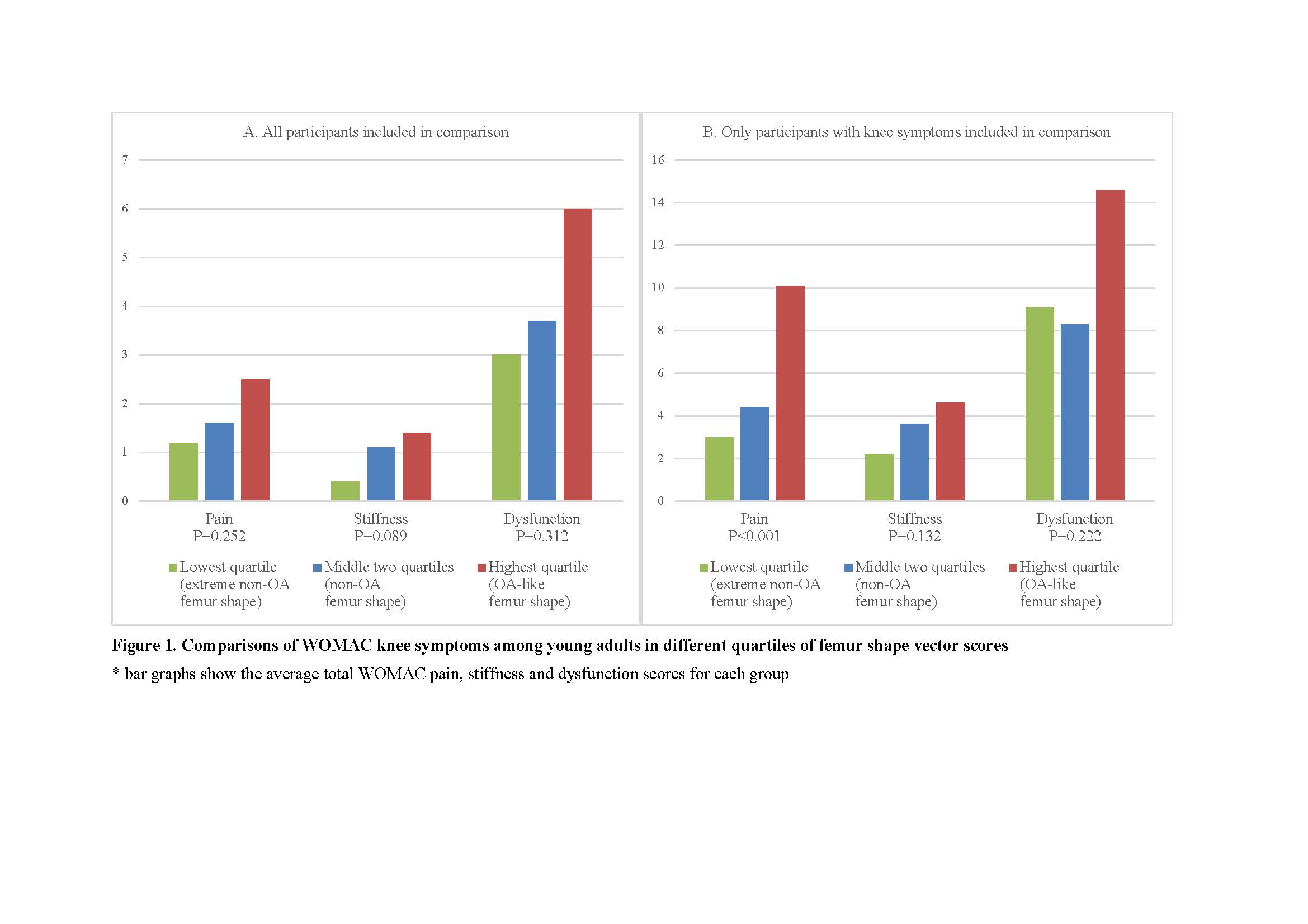

Figure1

Figure 1. Comparisons of WOMAC knee symptoms among young adults in different quartiles of femur shape vector scores


Disclosure: M. Vo, None; M. Bowes, Imorphics (Stryker), 3, OrthoTrophix, 9; A. Venn, None; F. Cicuttini, None; L. March, BMS, 8, Janssen, 8; T. Dwyer, None; T. Meng, None; M. Cross, None; G. Jones, None; S. Balogun, None; C. Ding, None; B. Antony, None.

To cite this abstract in AMA style:

Vo M, Bowes M, Venn A, Cicuttini F, March L, Dwyer T, Meng T, Cross M, Jones G, Balogun S, Ding C, Antony B. Clinical Significance of Magnetic Resonance Imaging Derived Femur Bone Shape in Young Adults [abstract]. Arthritis Rheumatol. 2019; 71 (suppl 10). https://acrabstracts.org/abstract/clinical-significance-of-magnetic-resonance-imaging-derived-femur-bone-shape-in-young-adults/. Accessed .
  • Tweet
  • Click to email a link to a friend (Opens in new window) Email
  • Click to print (Opens in new window) Print

« Back to 2019 ACR/ARP Annual Meeting

ACR Meeting Abstracts - https://acrabstracts.org/abstract/clinical-significance-of-magnetic-resonance-imaging-derived-femur-bone-shape-in-young-adults/

Advanced Search

Your Favorites

You can save and print a list of your favorite abstracts during your browser session by clicking the “Favorite” button at the bottom of any abstract. View your favorites »

All abstracts accepted to ACR Convergence are under media embargo once the ACR has notified presenters of their abstract’s acceptance. They may be presented at other meetings or published as manuscripts after this time but should not be discussed in non-scholarly venues or outlets. The following embargo policies are strictly enforced by the ACR.

Accepted abstracts are made available to the public online in advance of the meeting and are published in a special online supplement of our scientific journal, Arthritis & Rheumatology. Information contained in those abstracts may not be released until the abstracts appear online. In an exception to the media embargo, academic institutions, private organizations, and companies with products whose value may be influenced by information contained in an abstract may issue a press release to coincide with the availability of an ACR abstract on the ACR website. However, the ACR continues to require that information that goes beyond that contained in the abstract (e.g., discussion of the abstract done as part of editorial news coverage) is under media embargo until 10:00 AM ET on November 14, 2024. Journalists with access to embargoed information cannot release articles or editorial news coverage before this time. Editorial news coverage is considered original articles/videos developed by employed journalists to report facts, commentary, and subject matter expert quotes in a narrative form using a variety of sources (e.g., research, announcements, press releases, events, etc.).

Violation of this policy may result in the abstract being withdrawn from the meeting and other measures deemed appropriate. Authors are responsible for notifying colleagues, institutions, communications firms, and all other stakeholders related to the development or promotion of the abstract about this policy. If you have questions about the ACR abstract embargo policy, please contact ACR abstracts staff at [email protected].

Wiley

  • Online Journal
  • Privacy Policy
  • Permissions Policies
  • Cookie Preferences

© Copyright 2025 American College of Rheumatology