Session Information
Date: Wednesday, November 8, 2017
Title: Imaging of Rheumatic Diseases II: Focus on Rheumatoid Arthritis and Systemic Sclerosis
Session Type: ACR Concurrent Abstract Session
Session Time: 11:00AM-12:30PM
Clinical Correlate of Synovial Proliferation in Early Rheumatoid Arthritis
Background/Purpose: To determine whether semi-quantitative, quantitative assessment of synovitis severity or synovial perfusion data correlates best with clinical symptoms and inflammatory markers.
Methods: 116 patients (88 females, 28 males, mean age, 53±13 years) with early (i.e. symptoms < 24 months) RA underwent 3T dynamic contrast-enhanced (DCE) MRI of the most symptomatic wrist. Sequences obtained were: fat-saturated T1-weighted axial; fat-saturated T2-weighted coronal; T1-weighted coronal and fat-saturated post-contrast T1-weighted axial.
Analyses undertaken included:
1. Semi-quantitative grading of (a) synovial proliferation (RAMRIS) and (b) tenosynovitis.
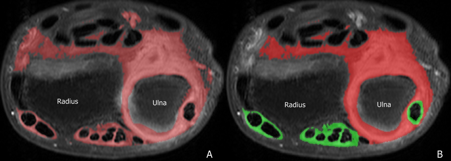
2. Quantitative measurement of enhancing synovial volume using segmentation method (cm3) (Fig 1).
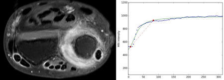
3. Maximum enhancement (ME) and enhancement slope (ES) of enhancing synovium derived from time-intensity curves of DCE MRI data (Fig 2).
4. Clinical assessment (Simple Disease Activity Index) and inflammatory markers (erythrocyte sedimentation rate, C-reactive protein).
Clinical evaluation included a determination of morning stiffness (minutes), pain score (0-10), disease activity by the Simple Disease Activity Index (which reflects the number of swollen ± tender joints) and serological inflammatory markers (ESR, CRP).
Results: Synovitis and tenosynovitis was present in 113 (97%) and 81 (70%) of the 116 wrists examined. Tenosynovitis was associated with more severe synovial proliferation. Regarding clinical correlation, quantitative parameters correlated much better with patient symptoms than semi-quantitative parameters. Morning stiffness and pain correlated with total synovial and tenosynovial volume (r=0.407, p=0.028) but not with RAMRIS (r=0.165, p=0.196). SDAI best correlated with ME and ES (r=0.502, p<0.001). ESR (r=0.380, p=0.002) and CRP (r=0.469, p<0.001) only correlated with ES.
Conclusion: Quantitative correlated with patient symptoms and serological inflammatory markers better than semi-quantitative parameters. Synovial volume was more related to morning stiffness and pain while perfusion parameters were more related to disease activity and inflammation.
Fig 1. Automated segmentation of enhancing synovium of the distal radioulnar joint (A) and following manual removal of adjacent vasculature (B). Joint synovial proliferation is seen as red, tenosynovial proliferation as green.
Fig 2. ERA patient (SDAI=40.46) with severe synovitis. The DCE perfusion curve was fit and Emax = 102.37, Eslope = 526.08 respectively.
To cite this abstract in AMA style:
XIAO F, YUE J, Mak Q, Tam LS, Griffith JF. Clinical Correlate of Synovial Proliferation in Early Rheumatoid Arthritis [abstract]. Arthritis Rheumatol. 2017; 69 (suppl 10). https://acrabstracts.org/abstract/clinical-correlate-of-synovial-proliferation-in-early-rheumatoid-arthritis/. Accessed .« Back to 2017 ACR/ARHP Annual Meeting
ACR Meeting Abstracts - https://acrabstracts.org/abstract/clinical-correlate-of-synovial-proliferation-in-early-rheumatoid-arthritis/


