Session Information
Session Type: Abstract Submissions (ACR)
Background/Purpose
CD4+ T cells and the Th1 and Th17 subsets in particular play a pivotal role in SLE. Regulatory T cells (Treg) are essential for maintaining peripheral tolerance, but their number and function in SLE is decreased. We herein characterize CD4+T cell and Treg homeostasis and severity of organ involvement in a murine model of SLE and analyze the effects of in vitro –induced iTreg.
Methods
Mice were injected i.p. with 0.5ml of pristane or PBS as control and killed after 8 months. Na•ve CD4+ thymocytes were cultured under specific conditions and tested for CD4+Foxp3+ expression by FACS. Cell suspensions with >80% purity for CD4+FoxP3+ iTreg were injected intravenously either once at start of experiments (iTreg-boost) or monthly (1xiTreg-repeated).
Animals were monitored for clinical signs of arthritis and, in order to analyze and compare disease severity, histological features of arthritis and pneumonitis were quantified by an image analysis system. Lungs were scored for the severity of perivascular inflammation by analyzing the numbers of affected vessels and the area of the inflammatory infiltrate.
Lymphocytes were isolated from granulomas (intraperitoneal ectopic lymphoid tissue), lymph nodes (LN) and spleens and were analyzed separately by FACS. For analyzes of the Th1, 2 and 17 subsets, cells were restimulated in vivo plate bound with anti-CD3 and anti-CD28abs.
Results
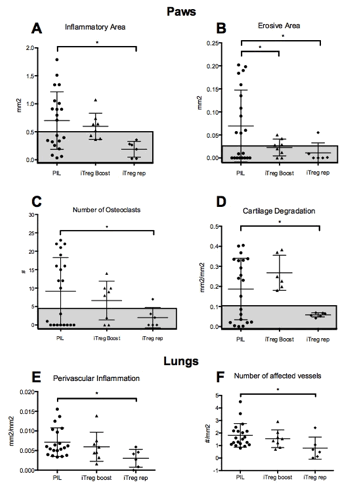
PIL mice presented with involvement of inner organs, most frequently affected were the lungs (100% pneumonitis); 52% of PIL developed arthritis, both clinically and histologically. Monthly iTreg-injection significantly decreased clinical signs of arthritis and histological lung and joint parameters. 66% of treated mice did not show any signs of arthritis at all. (Fig. 1A-F)
The iTreg-boost did not prevent joint manifestations or pneumonitis, but appeared to have a retarding effect (Fig.1A-F) indicated by a delayed onset of clinical symptoms and by a significantly decreased erosive area at the end of observation (Fig.1B).
Intraperitoneal granuloma typical for PIL appeared to be the hotspots of inflammation showing a significantly elevated Teff/Treg ratio of 1.3. Upon re-stimulation, CD4+ cells showed a pronounced Th1 response (27% IFNγ producers) compared to cells from LN and spleens from both PIL and HC (with Th1 percentages ranging from 9-16%). In addition, frequencies of Th2 and Th17 cells were elevated in PIL, again with the highest yield in granuloma. The repeated application of iTreg reduced the Teff/Treg ratio in PIL granuloma to 0.7.
Conclusion
Repeated injections of iTregs reduce severity of pneumonitis and arthritis as well as the Teff/Treg ratio. A single injection of iTregs is not effective, but appears to retard onset of symptoms and progression of arthritis and pneumonitis. Thus, iTreg have significant effects on lupus symptoms, which may have consequence for future therapeutic considerations.
Disclosure:
H. Leiss,
None;
B. Schwarzecker,
None;
I. Gessl,
None;
A. Puchner,
None;
B. Niederreiter,
None;
C. W. Steiner,
None;
J. Smolen,
None;
G. H. Stummvoll,
None.
« Back to 2014 ACR/ARHP Annual Meeting
ACR Meeting Abstracts - https://acrabstracts.org/abstract/characterization-of-cd4-t-cell-response-and-effects-of-regulatory-t-cells-in-pristane-induced-lupus-pil/

